Contour of lingual surface in lower complete denture formed by polished surface impression
- Affiliations
-
- 1Department of Prosthodontics, School of Dentistry, Chosun University, Gwangju, Republic of Korea. jhajung@chosun.ac.kr
- KMID: 2382593
- DOI: http://doi.org/10.4047/jap.2016.8.6.472
Abstract
- PURPOSE
The aim of this study was to analyze the shapes of lingual polished surfaces in lower complete dentures formed by polished surface impressions and to provide reference data for use when manufacturing edentulous trays and lower complete dentures.
MATERIALS AND METHODS
Twenty-six patients with mandibular edentulism were studied. After lower wax dentures were fabricated, wax was removed from the lingual side of the wax denture and a lingual polished surface impression was obtained with tissue conditioner. The definitive denture was scanned with a three-dimensional scanner, and scanned images were obtained. At the cross-sections of the lingual frenum, lateral incisors, first premolars, first molars, and anterior border of the retromolar pads, three points were marked and eight measurements were taken. The Kruskal-Wallis test and a post hoc analysis with the Mann-Whitney test were performed.
RESULTS
Each patient showed similar values for the same areas on the left and right sides without a statistically significant difference. The height of the contour of the lingual polished surface at the lingual frenum was halfway between the occlusal plane and lingual border, it moved gradually in a downward direction. The angle from the occlusal plane to the height of the contour of the lingual polished surface was increased as it progressed from the lingual frenum towards the retromolar pads.
CONCLUSION
The shape of the mandibular lingual polished surface was convex at the lingual frenum, lateral incisors and gradually flattened towards the first molars and retromolar pads.
Figure
-
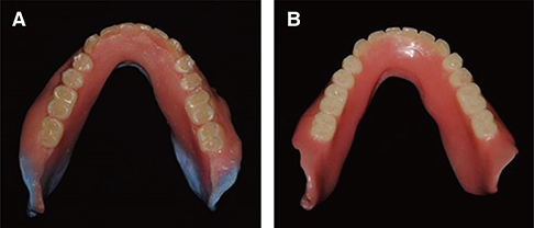
Fig. 1 (A) Lower wax denture formed by polished surface impression using a tissue conditioner, (B) Definitive lower compete denture.
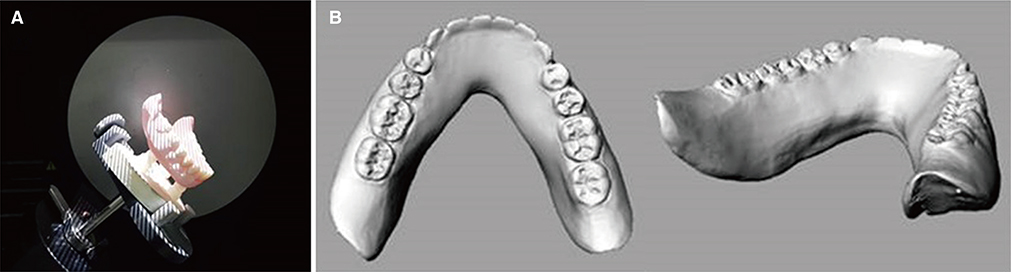
Fig. 2 (A) Scanning of the definitive denture, (B) Scanned images of definitive denture.
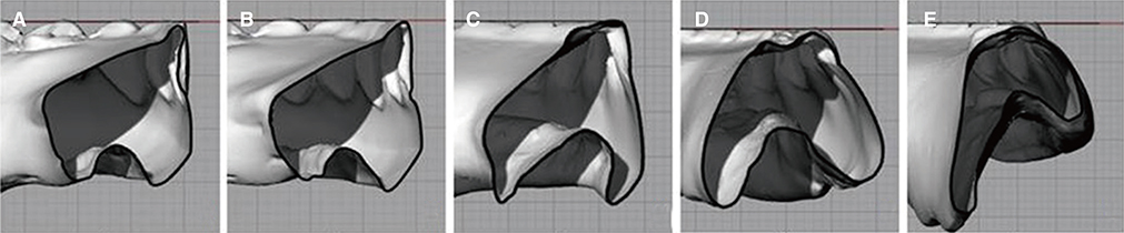
Fig. 3 Cross sections of each region. (A) lingual frenum, (B) lateral incisor, (C) first premolar, (D) first molar, (E) anterior border of retromolar pad.

Fig. 4 Reference points and measurements at the sectioned lingual polished surface. (a) the lingual margin of the artificial teeth, (b) the height of the contour at the lingual polished surface, (c) the most inferior lingual border, (A) the perpendicular distance between the a-c line and b, (B) the vertical distance from the occlusal plane to b, (C) the vertical distance between b and c, (D) the vertical distance from the occlusal plane to c, (E) the ratio of C to D, (F) the angle formed by the a-b line and the occlusal plane, (G) the angle formed by the b-c line and the occlusal plane, (H) the angle formed by the a-b line and the b-c line.
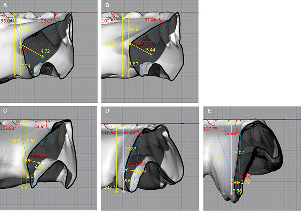
Fig. 5 Contour and mean values of each region. (A) lingual frenum, (B) lateral incisor, (C) first premolar, (D) first molar, (E) anterior border of retromolar pad.
Cited by 1 articles
-
Comparison of different impression techniques for edentulous jaws using three-dimensional analysis
Sua Jung, Chan Park, Hong-So Yang, Hyun-Pil Lim, Kwi-Dug Yun, Zhai Ying, Sang-Won Park
J Adv Prosthodont. 2019;11(3):179-186. doi: 10.4047/jap.2019.11.3.179.
Reference
-
1. Jacobson TE, Krol AJ. A contemporary review of the factors involved in complete denture retention, stability, and support. Part I: retention. J Prosthet Dent. 1983; 49:5–15.2. Beresin VE, Schiesser FJ. The neutral zone in complete dentures. J Prosthet Dent. 1976; 36:356–367.3. Bohnenkamp DM, Garcia LT. Phonetics and tongue position to improve mandibular denture retention: a clinical report. J Prosthet Dent. 2007; 98:344–347.4. Brill N, Tryde G, Cantor R. The dynamic nature of the lower denture space. J Prosthet Dent. 1965; 15:401–418.5. Shanahan TEJ. Stabilizing lower dentures on unfavorable ridges. J Prosthet Dent. 1962; 12:420–424.6. Srivastava V, Gupta NK, Tandan A, Kaira LS, Chopra D. The neutral zone: Concept and technique. J Orofac Res. 2012; 2:42–47.7. Fish EW. Principles of full denture prosthesis. . London: John Bale, Sons&Danielsson, Ltd;1933. p. 1–8.8. Neill DJ, Glaysher JK. Identifying the denture space. J Oral Rehabil. 1982; 9:259–277.9. Raybin NH. The polished surface of complete dentures. J Prosthet Dent. 1963; 13:236–239.10. Tuckfield WJ. The problem of the mandibular denture. Dent J Aust. 1951; 23:331–354.11. Tillman EJ. Removable partial upper and complete lower dentures. J Prosthet Dent. 1961; 11:1098–1104.12. Kwon KR, Kim YS, Kim CH, Kim HJ, Moon HS, Park SW, Park CJ, Song KY, Lee JS, Lee CH, Lim YJ, Jung MK, Chung CH, Jung CM, Cho IH, Cho HW, Choi DK, Han JH. Prosthodontic treatment for edentulous patient. Seoul: Shinhung Internarional, Inc;2007. p. 323.13. Sussman BA. Procedures in complete denture prosthesis. J Prosthet Dent. 1960; 10:1011–1021.14. Block LS. Common factors in complete denture prosthetics. J Prosthet Dent. 1953; 3:736–746.15. Starcke EN Jr. The contours of polished surfaces of complete dentures: a review of the literature. J Am Dent Assoc. 1970; 81:155–160.16. Cagna DR, Massad JJ, Schiesser FJ. The neutral zone revisited: from historical concepts to modern application. J Prosthet Dent. 2009; 101:405–412.17. Kolb HR. Variable denture-limiting structures of the edentulous mouth. II. Mandibular border areas. J Prosthet Dent. 1966; 16:202–212.
- Full Text Links
-
- Actions
-
Cited
- CITED
-
- Close
- Share
-
- Similar articles
-
- Complete denture making in a patient of partial glossectomy using polished surface impression taking and direct metal laser sintering method: A case report
- Using the neutral zone for a patient with bone resorption: a case report
- Neutral zone approach and external impression for rehabilitation of severely atrophic maxillary and mandibular ridges: a case report
- A morphologic characteristics study on crown of lingual surface with normal occlusion in Korean adults
- Tensile bond strength of four denture resins to porcelain teeth with different surface treatment
