J Pathol Transl Med.
2015 May;49(3):267-269. 10.4132/jptm.2015.03.20.
Traumatic Bowel Perforation and Inguinal Hernia Masking a Mesenteric Calcifying Fibrous Tumor
- Affiliations
-
- 1Department of Pathology, Kangbuk Samsung Hospital, Sungkyunkwan University School of Medicine, Seoul, Korea. idavid.kim@samsung.com
- 2Department of Pathology, Hallym University Sacred Heart Hospital, Hallym University College of Medicine, Anyang, Korea.
- KMID: 2381387
- DOI: http://doi.org/10.4132/jptm.2015.03.20
Abstract
- No abstract available.
MeSH Terms
Figure
-

Fig. 1. Abdominal contrast-enhanced computed tomography scan reveals diffuse enhancing wall thickening (arrow) without an obvious mass-like lesion.
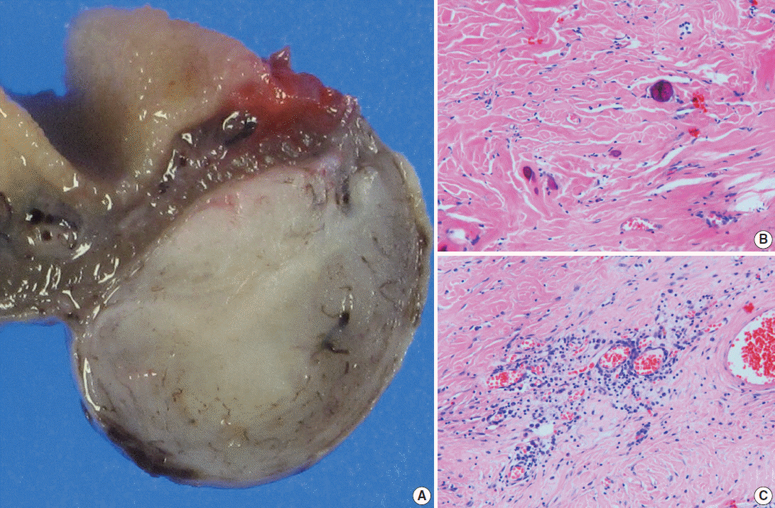
Fig. 2. (A) Viewed grossly, a well-demarcated gray-brown firm solid mass is confined to the mesenteric fat (right). (B) Microscopically, there are dispersed sparse spindle cells and occasional dystrophic calcification among thick wavy collagen bundles (left upper). (C) Patchy lymphoplasmacytic infiltrations are found throughout the tumor.
Reference
-
1. Emanuel P, Qin L, Harpaz N. Calcifying fibrous tumor of small intestine. Ann Diagn Pathol. 2008; 12:138–41.
Article2. Ben-Izhak O, Itin L, Feuchtwanger Z, Lifschitz-Mercer B, Czernobilsky B. Calcifying fibrous pseudotumor of mesentery presenting with acute peritonitis: case report with immunohistochemical study and review of literature. Int J Surg Pathol. 2001; 9:249–53.3. Giardino AA, Ramaiya NH, Shinagare AB, Jagannathan JP, Stachler MD, Raut CP. Case report: Calcifying fibrous tumor presenting as an asymptomatic pelvic mass. Indian J Radiol Imaging. 2011; 21:306–8.
Article4. Van Dorpe J, Ectors N, Geboes K, D’Hoore A, Sciot R. Is calcifying fibrous pseudotumor a late sclerosing stage of inflammatory myofibroblastic tumor? Am J Surg Pathol. 1999; 23:329–35.
Article5. Pomplun S, Goldstraw P, Davies SE, Burke MM, Nicholson AG. Calcifying fibrous pseudotumour arising within an inflammatory pseudotumour: evidence of progression from one lesion to the other? Histopathology. 2000; 37:380–2.
Article6. Sigel JE, Smith TA, Reith JD, Goldblum JR. Immunohistochemical analysis of anaplastic lymphoma kinase expression in deep soft tissue calcifying fibrous pseudotumor: evidence of a late sclerosing stage of inflammatory myofibroblastic tumor? Ann Diagn Pathol. 2001; 5:10–4.
Article7. Nascimento AF, Ruiz R, Hornick JL, Fletcher CD. Calcifying fibrous ‘pseudotumor’: clinicopathologic study of 15 cases and analysis of its relationship to inflammatory myofibroblastic tumor. Int J Surg Pathol. 2002; 10:189–96.8. Kuo TT, Chen TC, Lee LY. Sclerosing angiomatoid nodular transformation of the spleen (SANT): clinicopathological study of 10 cases with or without abdominal disseminated calcifying fibrous tumors, and the presence of a significant number of IgG4+ plasma cells. Pathol Int. 2009; 59:844–50.
Article9. Agaimy A, Bihl MP, Tornillo L, Wünsch PH, Hartmann A, Michal M. Calcifying fibrous tumor of the stomach: clinicopathologic and molecular study of seven cases with literature review and reappraisal of histogenesis. Am J Surg Pathol. 2010; 34:271–8.
Article10. Larson BK, Balzer B, Goldwasser J, Dhall D. Calcifying fibrous tumor: an unrecognized IgG4: related disease? APMIS. 2015; 123:72–6.
- Full Text Links
-
- Actions
-
Cited
- CITED
-
- Close
- Share
-
- Similar articles
-
- Obturator Hernia Which was Combined with Inguinal Hernia and Hiatal Hernia
- Delayed presenting traumatic diaphragmatic hernia: four case reports
- A Case of Functional Testicular Torsion Caused by Incarcerated Inguinal Hernia in a Newborn
- Non-traumatic Small Bowel Perforation: Comparisons of the Clinical Features of 20 Years Ago with Present
- Amyand's Hernia with Periappendicular Abscess
