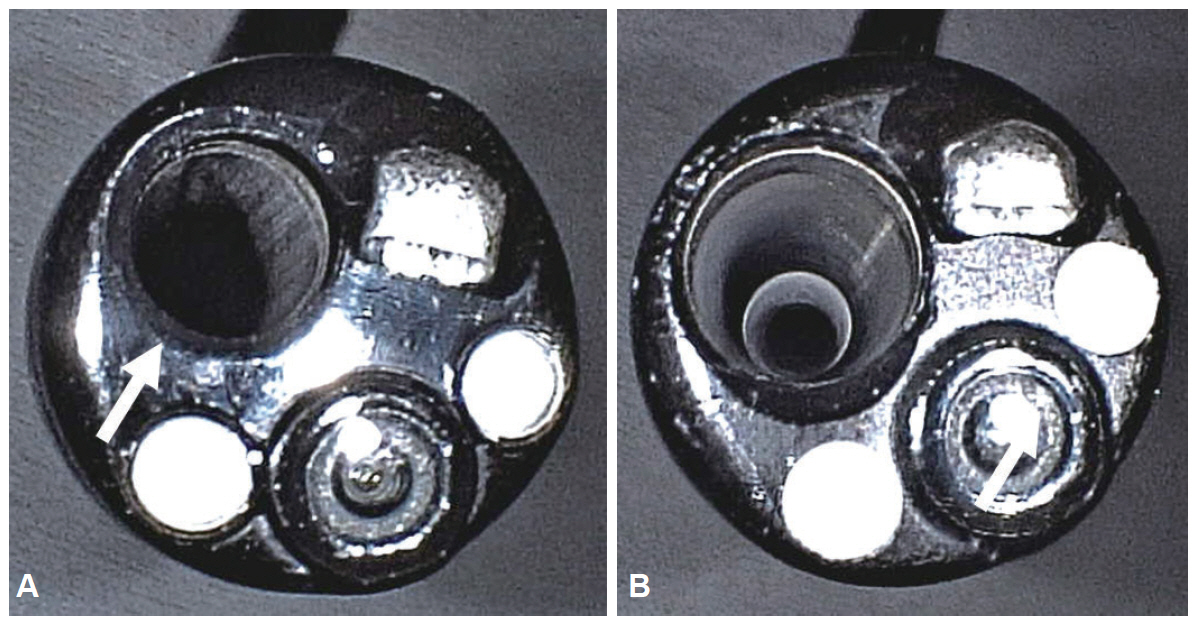
Clin Endosc.
2015 Nov;48(6):516-521. 10.5946/ce.2015.48.6.516.
An Ultrathin Endoscope with a 2.4-mm Working Channel Shortens the Esophagogastroduodenoscopy Time by Shortening the Suction Time
- Affiliations
-
- 1Department of Gastroenterology, Shinozaki Medical Clinic, Tochigi, Japan.
- 2Division of Gastroenterology, Department of Medicine, Japan. ireef@jichi.ac.jp
- 3Department of Surgery, Jichi Medical University School of Medicine, Shimotsuke, Japan.
- KMID: 2380409
- DOI: http://doi.org/10.5946/ce.2015.48.6.516
Abstract
- BACKGROUND/AIMS
Poor suction ability through a narrow working channel prolongs esophagogastroduodenoscopy (EGD). The aim of this study was to evaluate suction with a new ultrathin endoscope (EG-580NW2; Fujifilm Corp.) having a 2.4-mm working channel in clinical practice.
METHODS
To evaluate in vitro suction, 200 mL water was suctioned and the suction time was measured. The clinical data of 117 patients who underwent EGD were retrospectively reviewed on the basis of recorded video, and the suction time was measured by using a stopwatch.
RESULTS
In vitro, the suction time with the EG-580NW2 endoscope was significantly shorter than that with the use of an ultrathin endoscope with a 2.0-mm working channel (EG-580NW; mean +/- standard deviation, 22.7+/-1.1 seconds vs. 34.7+/-2.2 seconds; p<0.001). We analyzed the total time and the suction time for routine EGD in 117 patients (50 in the EG-580NW2 group and 67 in the EG-580NW group). In the EG-580NW2 group, the total time for EGD was significantly shorter than that in the EG-580NW group (275.3+/-42.0 seconds vs. 300.6+/-46.5 seconds, p=0.003). In the EG-580NW2 group, the suction time was significantly shorter than that in the EG-580NW group (19.2+/-7.6 seconds vs. 38.0+/-15.9 seconds, p<0.001).
CONCLUSIONS
An ultrathin endoscope with a 2.4-mm working channel considerably shortens the routine EGD time by shortening the suction time, in comparison with an endoscope with a 2.0-mm working channel.
Keyword
MeSH Terms
Figure
Reference
-
1. Kadayifci A, Atar M, Parlar S, Balkan A, Koruk I, Koruk M. Transnasal endoscopy is preferred by transoral endoscopy experienced patients. J Gastrointestin Liver Dis. 2014; 23:27–31.
Article2. Murata A, Akahoshi K, Motomura Y, et al. Prospective comparative study on the acceptability of unsedated transnasal endoscopy in younger versus older patients. J Clin Gastroenterol. 2008; 42:965–968.
Article3. Atar M, Kadayifci A. Transnasal endoscopy: technical considerations, advantages and limitations. World J Gastrointest Endosc. 2014; 6:41–48.
Article4. Trevisani L, Cifalà V, Sartori S, Gilli G, Matarese G, Abbasciano V. Unsedated ultrathin upper endoscopy is better than conventional endoscopy in routine outpatient gastroenterology practice: a randomized trial. World J Gastroenterol. 2007; 13:906–911.
Article5. Dumortier J, Josso C, Roman S, et al. Prospective evaluation of a new ultrathin one-plane bending videoendoscope for transnasal EGD: a comparative study on performance and tolerance. Gastrointest Endosc. 2007; 66:13–19.
Article6. Garcia RT, Cello JP, Nguyen MH, et al. Unsedated ultrathin EGD is well accepted when compared with conventional sedated EGD: a multicenter randomized trial. Gastroenterology. 2003; 125:1606–1612.
Article7. ASGE Technology Committee, Rodriguez SA, Banerjee S, et al. Ultrathin endoscopes. Gastrointest Endosc. 2010; 71:893–898.
Article
- Full Text Links
- Actions
-
Cited
- CITED
-
- Close
- Share
- Similar articles
-
- An Ultrathin Endoscope with a 2.4-mm Working Channel Shortens the Esophagogastroduodenoscopy Time by Shortening the Suction Time
- Why the Large Working Channel Uniportal Endoscope is Better for Patients With Obesity Than the Unilateral Biportal Endoscope at Lower Lumbar Levels: A Technical Note
- Ultrathin Endoscope-Assisted Method for the Management of Upper Gastrointestinal Obstruction to Avoid Technical Failure
- Placement of a Self-Expanding Metal Stent to Treat Esophagogastric Benign Anastomotic Stricture via Retroflexed Ultrathin Endoscopy: A Case Report with a Video
- A Comparison of the Opened Versus Closed-System of Suctioning : In Oxygen Saturation, Vital Signs and Suction Time


