Cancer Res Treat.
2015 Jan;47(1):120-125. 10.4143/crt.2013.137.
MET-Amplified Intramucosal Gastric Cancer Widely Metastatic after Complete Endoscopic Submucosal Dissection
- Affiliations
-
- 1Department of Pathology, Ajou University School of Medicine, Suwon, Korea. ybkim@ajou.ac.kr
- 2Department of Radiology, Ajou University School of Medicine, Suwon, Korea.
- 3Department of Gastroenterology, Ajou University School of Medicine, Suwon, Korea.
- 4Department of Nuclear Medicine and Molecular Imaging, Ajou University School of Medicine, Suwon, Korea.
- KMID: 2380398
- DOI: http://doi.org/10.4143/crt.2013.137
Abstract
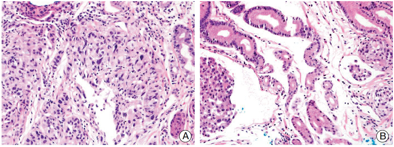
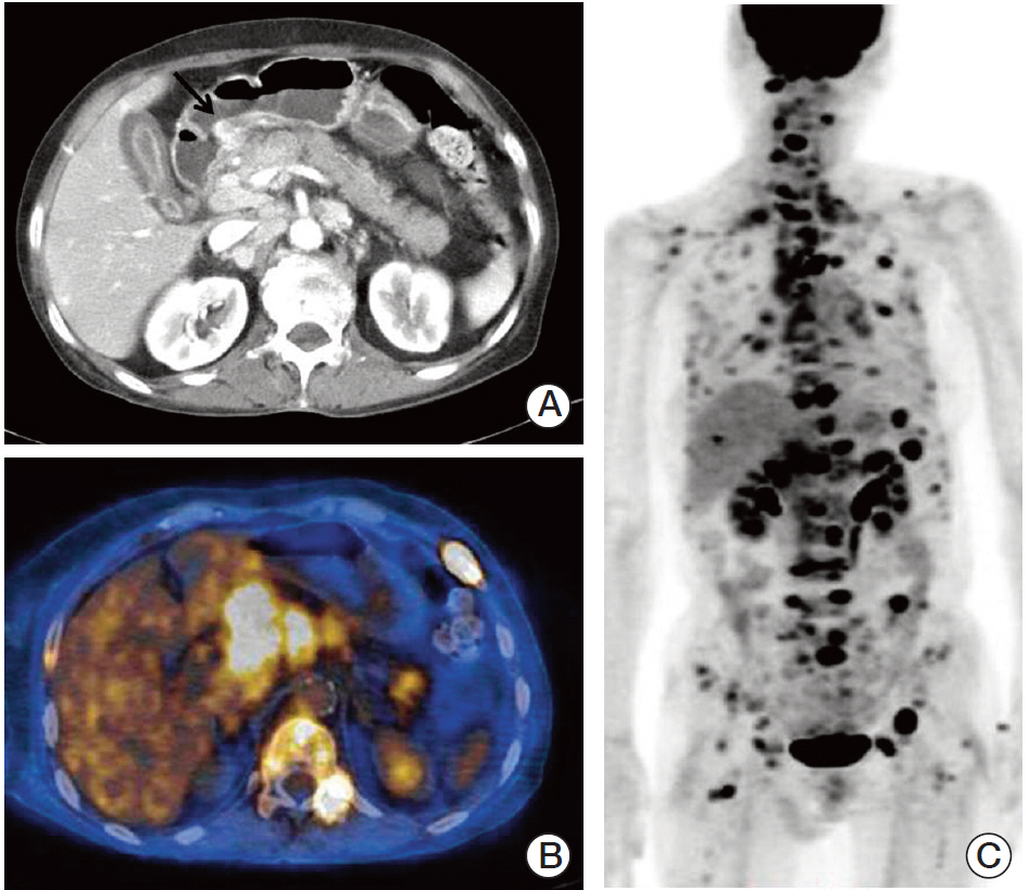
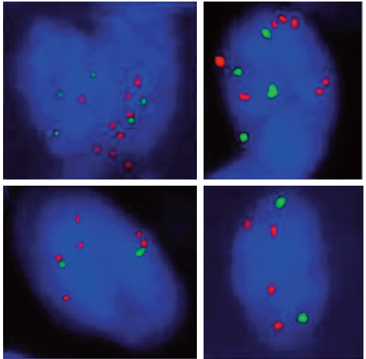
- Intramucosal gastric cancer (IGC) is associated with a very low risk of lymph node metastasis; thus it is the main candidate for minimally invasive surgical procedures, such as endoscopic submucosal dissection (ESD). Herein, we document an extraordinary case of IGC, which showed a very aggressive clinical course. A 66-year-old female underwent ESD for early gastric cancer. Histologically, the tumor consisted mainly of moderately differentiated adenocarcinoma measuring 1.6 cm in diameter, and the tumor was confined to the mucosa. Despite annual esophagogastroduodenoscopic follow-up, the tumor recurred, with wide metastasis to multiple lymph nodes and bones throughout the body after three years. Fluorescence in situ hybridization study demonstrated MET gene amplification as well as low grade polysomy 7 in both original and recurrent tumors. The clinical characteristics of metastatic IGCs and the implication of MET amplification are discussed.
Keyword
MeSH Terms
Figure
Reference
-
References
1. Angeles-Angeles A, Candanedo-Gonzalez F, Gamboa-Dominguez A, Larriva-Sahd J. A clinicopathologic variant of intramucosal early gastric cancer with widespread dissemination: report of three cases. J Clin Gastroenterol. 1998; 27:173–7.2. Hanaoka N, Tanabe S, Higuchi K, Sasaki T, Nakatani K, Ishido K, et al. A rare case of histologically mixed-type intramucosal gastric cancer accompanied by nodal recurrence and liver metastasis after endoscopic submucosal dissection. Gastrointest Endosc. 2009; 69(3 Pt 1):588–90.
Article3. Oya M, Yao T, Nagai E, Tsuneyoshi M. Metastasizing intramucosal gastric carcinomas: well differentiated type and proliferative activity using proliferative cell nuclear antigen and Ki-67. Cancer. 1995; 75:926–35.
Article4. Song SY, Park S, Kim S, Son HJ, Rhee JC. Characteristics of intramucosal gastric carcinoma with lymph node metastatic disease. Histopathology. 2004; 44:437–44.
Article5. Graziano F, Galluccio N, Lorenzini P, Ruzzo A, Canestrari E, D'Emidio S, et al. Genetic activation of the MET pathway and prognosis of patients with high-risk, radically resected gastric cancer. J Clin Oncol. 2011; 29:4789–95.
Article6. Lennerz JK, Kwak EL, Ackerman A, Michael M, Fox SB, Bergethon K, et al. MET amplification identifies a small and aggressive subgroup of esophagogastric adenocarcinoma with evidence of responsiveness to crizotinib. J Clin Oncol. 2011; 29:4803–10.
Article7. Oh HS, Eom DW, Kang GH, Ahn YC, Lee SJ, Kim JH, et al. Prognostic implications of EGFR and HER-2 alteration assessed by immunohistochemistry and silver in situ hybridization in gastric cancer patients following curative resection. Gastric Cancer. 2014; 17:402–11.
Article8. Bang YJ, Van Cutsem E, Feyereislova A, Chung HC, Shen L, Sawaki A, et al. Trastuzumab in combination with chemotherapy versus chemotherapy alone for treatment of HER2-positive advanced gastric or gastro-oesophageal junction cancer (ToGA): a phase 3, open-label, randomised controlled trial. Lancet. 2010; 376:687–97.
Article9. Jung EJ, Jung EJ, Min SY, Kim MA, Kim WH. Fibroblast growth factor receptor 2 gene amplification status and its clinicopathologic significance in gastric carcinoma. Hum Pathol. 2012; 43:1559–66.
Article10. Shi J, Yao D, Liu W, Wang N, Lv H, Zhang G, et al. Highly frequent PIK3CA amplification is associated with poor prognosis in gastric cancer. BMC Cancer. 2012; 12:50.
Article11. Korenaga D, Haraguchi M, Tsujitani S, Okamura T, Tamada R, Sugimachi K. Clinicopathological features of mucosal carcinoma of the stomach with lymph node metastasis in eleven patients. Br J Surg. 1986; 73:431–3.
Article12. Kakushima N, Kamoshida T, Hirai S, Hotta S, Hirayama T, Yamada J, et al. Early gastric cancer with Krukenberg tumor and review of cases of intramucosal gastric cancers with Krukenberg tumor. J Gastroenterol. 2003; 38:1176–80.
Article13. Yamao T, Shirao K, Ono H, Kondo H, Saito D, Yamaguchi H, et al. Risk factors for lymph node metastasis from intramucosal gastric carcinoma. Cancer. 1996; 77:602–6.
Article14. Corso S, Comoglio PM, Giordano S. Cancer therapy: can the challenge be MET? Trends Mol Med. 2005; 11:284–92.
Article15. Lee J, Seo JW, Jun HJ, Ki CS, Park SH, Park YS, et al. Impact of MET amplification on gastric cancer: possible roles as a novel prognostic marker and a potential therapeutic target. Oncol Rep. 2011; 25:1517–24.
Article
- Full Text Links
- Actions
-
Cited
- CITED
-
- Close
- Share
- Similar articles
-
- Endoscopic Resection of Undifferentiated Early Gastric Cancer
- Endoscopic Treatment for Early Gastric Cancer
- Pathological Interpretation of Gastric Tumors in Endoscopic Submucosal Dissection
- Unusual Local Recurrence with Distant Metastasis after Successful Endoscopic Submucosal Dissection for Colorectal Mucosal Cancer
- Discrepancy between Clinical and Final Pathological Evaluation Findings in Early Gastric Cancer Patients Treated with Endoscopic Submucosal Dissection


