J Korean Soc Radiol.
2017 Jun;76(6):438-441. 10.3348/jksr.2017.76.6.438.
Percutaneous Biopsy of a Metastatic Common Iliac Lymph Node Using Hydrodissection and a Semi-Automated Biopsy Gun
- Affiliations
-
- 1Department of Radiology, Samsung Medical Center, Sungkyunkwan University School of Medicine, Seoul, Korea. rapark@skku.edu
- KMID: 2379328
- DOI: http://doi.org/10.3348/jksr.2017.76.6.438
Abstract
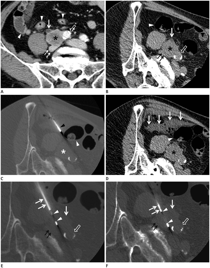
- Percutaneous biopsy is a less invasive technique for sampling the tissue than laparoscopic biopsy or exploratory laparotomy. However, it is difficult to perform biopsy of a deep-seated lesion because of the possibility of damage to the critical organs. Recently, we successfully performed CT-guided biopsy of a metastatic common iliac lymph node using hydrodissection and semi-automated biopsy devices. The purpose of this case report was to show how to perform hydrodissection and how to use a semi-automated gun for safe biopsy of a metastatic common iliac lymph node.
Figure
Reference
-
1. Chade DC, Shariat SF, Cronin AM, Savage CJ, Karnes RJ, Blute ML, et al. Salvage radical prostatectomy for radiation-recurrent prostate cancer: a multi-institutional collaboration. Eur Urol. 2011; 60:205–210.2. Farrell MA, Charboneau JW, Callstrom MR, Reading CC, Engen DE, Blute ML. Paranephric water instillation: a technique to prevent bowel injury during percutaneous renal radiofrequency ablation. AJR Am J Roentgenol. 2003; 181:1315–1317.3. Asvadi NH, Arellano RS. Hydrodissection-assisted image-guided percutaneous biopsy of abdominal and pelvic lesions: experience with seven patients. AJR Am J Roentgenol. 2015; 204:865–867.4. Park SY, Park BK, Kim CK, Kwon GY. Ultrasound-guided core biopsy of small renal masses: diagnostic rate and limitations. J Vasc Interv Radiol. 2013; 24:90–96.
- Full Text Links
- Actions
-
Cited
- CITED
-
- Close
- Share
- Similar articles
-
- Usefulness of US-Guided Automated Gun Biopsy of Neck Masses
- Ultrasound-Guided Renal Biopsy with Automated Biopsy Gun Technique: Efficacy and Complications
- US-Guided Percutaneous Renal Biopsy by Using 18 G Automatic Biopsy Gun in Diffuse Renal Disease
- CT-Guided Percutaneous Automated Gun Biopsy of Pulmonary Lesions: Complications and Diagnostic Accuracy
- Ultrasound Guided Biopsy of Malignant Focal Liver Lesions: Comparison of Automated Gun Biopsy with Fine Needle Aspiration Biopsy


