Ann Dermatol.
2017 Jun;29(3):363-364. 10.5021/ad.2017.29.3.363.
Papular Acantholytic Dyskeratosis of the Inguinal Area in a 49-Year-Old Man
- Affiliations
-
- 1Department of Dermatology, Ajou University School of Medicine, Suwon, Korea. maychan@ajou.ac.kr
- KMID: 2378544
- DOI: http://doi.org/10.5021/ad.2017.29.3.363
Abstract
- No abstract available.
MeSH Terms
Figure
-

Fig. 1 Multiple whitish pruritic papules with underlying erythematous patches were noted on the right (A) and left (B) inguinal areas. Steroid-induced erythematous atrophic patches were also found.
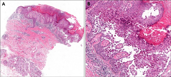
Fig. 2 Skin biopsy showed suprabasal cleft with villi, and numerous typical acantholytic dyskeratotic cells in the stratum corneum and spinosum. Hyperkeratosis and irregular acanthosis were also present in the epidermis, with a lymphocytic infiltration in the underlying dermis (H&E; A: ×40, B: ×200).
Reference
-
1. Knopp EA, Saraceni C, Moss J, McNiff JM, Choate KA. Somatic ATP2A2 mutation in a case of papular acantholytic dyskeratosis: mosaic Darier disease. J Cutan Pathol. 2015; 42:853–857.
Article2. Montis-Palos MC, Acebo-Mariñas E, Catón-Santarén B, Soloeta-Arechavala R. Papular acantholytic dermatosis in the genito-crural region: a localized form of Darier disease or Hailey-Hailey disease? Actas Dermosifiliogr. 2013; 104:170–172.
Article3. Dowd ML, Ansell LH, Husain S, Grossman ME. Papular acantholytic dyskeratosis of the genitocrural area: a rare unilateral asymptomatic intertrigo. JAAD Case Rep. 2016; 2:132–134.
Article4. Jue SJ, Kim JH, Ro YS, Lee CW, Kim JH. A case of papular acantholytic dermatosis localized to the anogenital area. Korean J Dermatol. 2002; 40:1121–1124.5. Lee JH, Kim YC, Lew W. A case of focal acantholytic dyskeratosis occurring on both the lip and the anal canal. Yonsei Med J. 2003; 44:166–168.
Article
- Full Text Links
-
- Actions
-
Cited
- CITED
-
- Close
- Share
-
- Similar articles
-
- A Case of Papular Acantholytic Dyskeratosis Treated with Carbon Dioxide Laser
- A Case of Focal Acantholytic Dyskeratosis Presenting as a Solitary Papule
- A Case of Focal Acantholytic Dyskeratosis Occurring on both the Lip and the Anal Canal
- Comments to "Localized Darier's Disease Mimicking Lichen Simplex Chronicus on the Back"
- A Case of Acantholytic Acanthoma
