Korean J Neurotrauma.
2015 Oct;11(2):70-74. 10.13004/kjnt.2015.11.2.70.
Clinical Features According to the Histological Types of the Outer Membrane of Chronic Subdural Hematoma
- Affiliations
-
- 1Department of Neurosurgery, Kangnam Sacred Heart Hospital, Hallym University College of Medicine, Seoul, Korea. nskch@hallym.or.kr
- 2Korea CFC Pathology Laboratory, Seongnam, Korea.
- KMID: 2378261
- DOI: http://doi.org/10.13004/kjnt.2015.11.2.70
Abstract
OBJECTIVE
The aim of our study was to classify the outer membrane of chronic subdural hematoma (CSDH) histologically and to determine the clinical and radiological meaning of the classified membranes.
METHODS
The outer membrane specimen of 31 patients who underwent surgery for CSDH were acquired in this study. The specimen was classified into four types and each were analyzed of the symptoms on the admission day and during the period from trauma to surgery. The radiological features such as subdural fluid density, Hounsfield number, thickness of the hematoma, and midline shift were analyzed.
RESULTS
There were 6% of type I, 29% of type II, 39% of type III, and 26% of type IV neomembranes. The cases of CSDH accompanied by neurologic deficit were highest from type IV of 63%, followed by type II with 56%. On the radiological findings such as Hounsfield unit, hematoma thickness and midline shift, only hematoma thickness between type II and III were statistically significant (p=0.021). The hematoma thickness and midline shift were greatest in type II. On computed tomography scans, the isodense, hyperdense and laminar type that shows the high recurrence rate formed 75% of type II and 67% of type IV while type III had the low possibility of recurrence rate (33%).
CONCLUSION
We have identified that the outer membrane have the tendency to develop from type I to IV in time while type II and type IV may have more risk of neurologic deficit and the high possibility of recurrence.
MeSH Terms
Figure
-
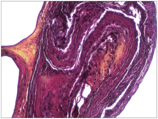
FIGURE 1 Photomicrograph of type I non-inflammatory membrane stained with hematoxylin and eosin and Elastica-van Gieson staining under light microscopy (original magnification, ×100).
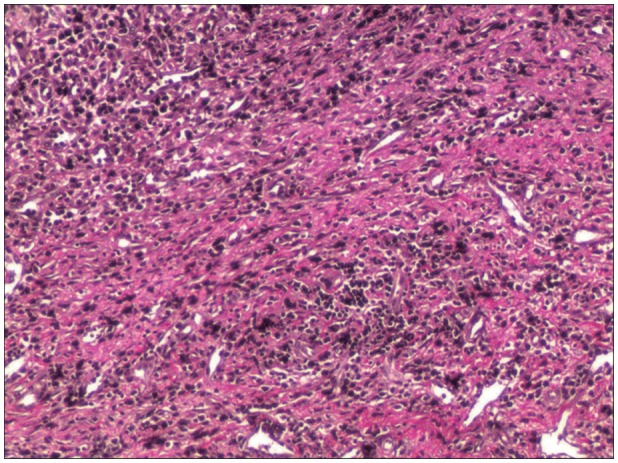
FIGURE 2 Photomicrograph of type II inflammatory membrane stained with hematoxylin and eosin and Elastica-van Gieson staining under light microscopy (original magnification, ×100).
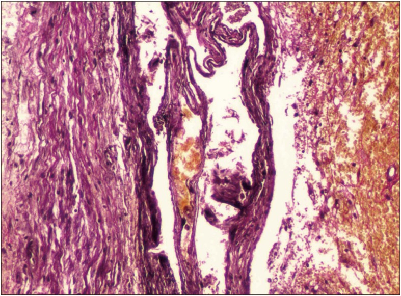
FIGURE 3 Photomicrograph of type III hemorrhagic inflammatory membrane stained with hematoxylin and eosin and Elastica-van Gieson staining under light microscopy (original magnification, ×100).
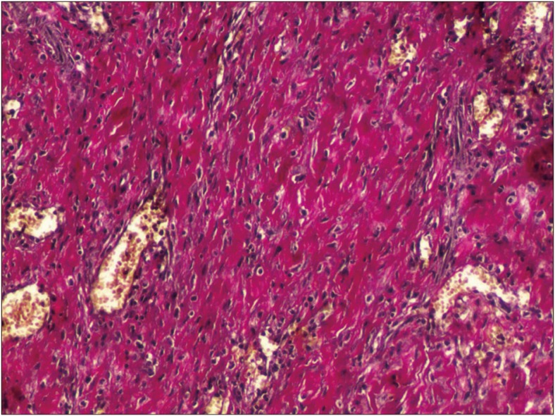
FIGURE 4 Photomicrograph of type IV scar inflammatory membrane stained with hematoxylin and eosin and Elastica-van Gieson staining under light microscopy (original magnification, ×100).
Reference
-
1. Ahn SY, Kim JH, Ha SK, Kim JH, Kwon TH, Park YK, et al. Clinical analysis of risk factors associated with the recurrence of chronic subdural hematoma. J Korean Neurotraumatol Soc. 2011; 7:68–73.
Article2. Friede RL. Incidence and distribution of neomembranes of dura mater. J Neurol Neurosurg Psychiatry. 1971; 34:439–446. PMID: 5096558.
Article3. Friede RL, Schachenmayr W. The origin ofsubdural neomembranes. II. Fine structural of neomembranes. Am J Pathol. 1978; 92:69–84. PMID: 686149.4. Gandhoke GS, Kaif M, Choi L, Williamson RW, Nakaji P. Histopathological features of the outer membrane of chronic subdural hematoma and correlation with clinical and radiological features. J Clin Neurosci. 2013; 20:1398–1401. PMID: 23916760.
Article5. Hara M, Tamaki M, Aoyagi M, Ohno K. Possible role of cyclooxygenase-2 in developing chronic subdural hematoma. J Med Dent Sci. 2009; 56:101–106. PMID: 20099472.6. Hong HJ, Kim YJ, Yi HJ, Ko Y, Oh SJ, Kim JM. Role of angiogenic growth factors and inflammatory cytokine on recurrence of chronic subdural hematoma. Surg Neurol. 2009; 71:161–165. discussion 165-166PMID: 18423527.
Article7. Ito H, Yamamoto S, Komai T, Mizukoshi H. Role of local hyperfibrinolysis in the etiology of chronic subdural hematoma. J Neurosurg. 1976; 45:26–31. PMID: 132513.
Article8. Jeong SI, Kim SO, Won YS, Kwon YJ, Choi CS. Clinical analysis of risk factors for recurrence in patients with chronic subdural hematoma undergoing burr hole trephination. Korean J Neurotrauma. 2014; 10:15–21.
Article9. Lee JK, Choi JH, Kim CH, Lee HK, Moon JG. Chronic subdural hematomas: a comparative study of three types of operative procedures. J Korean Neurosurg Soc. 2009; 46:210–214. PMID: 19844620.10. Lee KS, Bae WK, Bae HG, Doh JW, Yun IG. The computed tomographic attenuation and the age of subdural hematomas. J Korean Med Sci. 1997; 12:353–359. PMID: 9288636.
Article11. Nagahori T, Nishijima M, Takaku A. [Histological study of the outer membrane of chronic subdural hematoma: possible mechanism for expansion of hematoma cavity]. No Shinkei Geka. 1993; 21:697–701. PMID: 8361567.12. Park HR, Lee KS, Shim JJ, Yoon SM, Bae HG, Doh JW. Multiple densities of the chronic subdural hematoma in CT scans. J Korean Neurosurg Soc. 2013; 54:38–41. PMID: 24044079.
Article13. Sato S, Suzuki J. Ultrastructural observations of the capsule of chronic subdural hematoma in various clinical stages. J Neurosurg. 1975; 43:569–578. PMID: 1181389.
Article14. Schachenmayr W, Friede RL. The origin of subdural neomembranes. I. Fine structure of the dura-arachnoid interface in man. Am J Pathol. 1978; 92:53–68. PMID: 686148.15. Shono T, Inamura T, Morioka T, Matsumoto K, Suzuki SO, Ikezaki K, et al. Vascular endothelial growth factor in chronic subdural haematomas. J Clin Neurosci. 2001; 8:411–415. PMID: 11535006.
Article16. Stanišić M, Hald J, Rasmussen IA, Pripp AH, Ivanović J, Kolstad F, et al. Volume and densities of chronic subdural haematoma obtained from CT imaging as predictors of postoperative recurrence: a prospective study of 107 operated patients. Acta Neurochir (Wien). 2013; 155:323–333. discussion 333PMID: 23229873.
Article17. Suzuki K, Takano S, Nose T, Doi M, Ohashi N. Increased concentration of vascular endothelial growth factor (VEGF) in chronic subdural hematoma. J Trauma. 1999; 46:532–533. PMID: 10088869.18. Vaquero J, Zurita M, Cincu R. Vascular endothelial growth-permeability factor in granulation tissue of chronic subdural haematomas. Acta Neurochir (Wien). 2002; 144:343–346. discussion 347PMID: 12021880.
- Full Text Links
-
- Actions
-
Cited
- CITED
-
- Close
- Share
-
- Similar articles
-
- Histopathologic Study of the Outer Membrane of Chronic Subdural Hematoma Related to Membrane Permeability
- The Difference of Membrane Permeability of Chronic Subdural Hematoma Related to Attenuation on computerized Tomography
- Endovascular surgery for chronic subdural hematoma
- Chronic Subdural Hematoma Superimposed on Posttraumatic Subdural Hygroma: A Report of Three Cases
- Treatment of Chronic Subdural Hematoma with Arachnoid Cyst
