Importance of Hematoma Removal Ratio in Ruptured Middle Cerebral Artery Aneurysm Surgery with Intrasylvian Hematoma
- Affiliations
-
- 1Department of Neurosurgery, School of Medicine, Inha University, Incheon, Korea. nsshim60@gmail.com
- KMID: 2377621
- DOI: http://doi.org/10.7461/jcen.2017.19.1.5
Abstract
OBJECTIVE
Ruptured middle cerebral artery (MCA) aneurysm with intrasylvian hematoma usually accompanied by progressive cerebral swelling with poorer outcomes. The authors present characteristics and importance of intrasylvian hematoma removal in the aneurysm surgery.
MATERIALS AND METHODS
From 2012 February to 2014 March, 24 aneurysm surgeries for ruptured MCA aneurysms with intrasylvian hematoma were performed in the authors' clinic. The patients were classified according to three groups. Group A included patients who underwent decompressive craniectomy within a few days after aneurysm surgery due to progressive cerebral swelling, group B included patients for whom decompression was not necessary, and group C included patients who showed severe cerebral swelling on admission and decompressive craniectomy and aneurysm surgery in one stage.
RESULTS
The mean hematoma volume on admission was 28.56 mL, 24.96 mL, and 66.78 mL for groups A, B and C, respectively. Removal of a larger amount of hematoma was observed on postoperative computerized tomography scan in groups B and C (63.2% and 59.0%) compared with group A (33.4%). Although no statistical difference was found between group A and group B (p = 0.115), it tends to show the lesser amount of hematoma removed, the more likely cerebral swelling will progress.
CONCLUSION
The lesser amount of hematoma in ruptured MCA aneurysm with intrasylvian hematoma tends to show benign clinical course than larger amounts. But, even if the hematoma is not easily removed in the operation, we suggest the other procedures such as continuous external catheter drainage of hematoma to avoid unnecessary coagulation or brain retraction.
Keyword
MeSH Terms
Figure
-
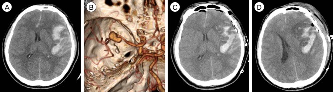
Fig. 1 CT and CT angiography in case 1. (A, B) Initial CT scans reveal characteristic findings of ruptured MCA aneurysm with intrasylvian hematoma. Hematoma volume was calculated as 17.6 mL. (C) Immediate postoperative CT finding of residual intrasylvian hematoma not sufficiently removed during aneurysm surgery. (D) Aggravated perihematoma swelling with midline sifting on POD 3. CT = computed tomography; MCA = middle cerebral artery; POD = post-operative day.
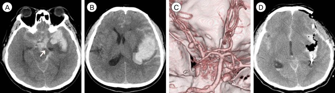
Fig. 2 CT and CT angiography in case 2. (A-C) SAH and intrasylvian hematoma from ruptured MCA aneurysm calculated as 64.9 mL. The arrow indicates uncal herniation. (D) Immediate postoperative CT reveals near total removal of intraysylvian hematoma. CT = computed tomography; SAH = subarachnoid hemorrhage; MCA = middle cerebral artery.
Reference
-
1. Başkaya MK, Menendez JA, Yüceer N, Polin RS, Nanda A. Results of surgical treatment of intrasylvian hematomas due to ruptured intracranial aneurysms. Clin Neurol Neurosurg. 2011; 4. 103(1):23–28.
Article2. Fisher CM, Kistler JP, Davis JM. Relation of cerebral vasospasm to subarachnoid hemorrhage visualized by computerized tomographic scanning. Neurosurgery. 1980; 1. 6(1):1–9. PMID: 7354892.
Article3. Jennett B, Snoek J, Bond MR, Brooks N. Disability after severe head injury: observations on the use of the Glasgow outcome scale. J Neurol Neurosurg Psychiatry. 1981; 4. 44(4):285–293. PMID: 6453957.
Article4. Jickling GC, Liu D, Stamova B, Ander BP, Zhan X, Lu A, et al. Hemorrhagic transformation after ischemic stroke in animals and humans. J Cereb Blood Flow Metab. 2014; 2. 34(2):185–199. PMID: 24281743.
Article5. Kopera M, Majchrzak H, Kaspera W. Prognostic factors in patients with intracerebral hematoma caused by ruptured middle cerebral artery aneurysm. Neurol Neurochir Pol. 1999; Mar-Apr. 33(2):389–401. PMID: 10463253.6. Kothari RU, Brott T, Broderick JP, Barsan WG, Sauerbeck LR, Zuccarello M, et al. The ABCs of measuring intracerebral hemorrhage volumes. Stroke. 1996; 8. 27(8):1304–1305. PMID: 8711791.
Article7. Pasqualin A, Bazzan A, Cavazzani P, Scienza R, Licata C, Da Pian R. Intracranial hematomas following aneurysmal rupture: experience with 309 cases. Surg Neurol. 1986; 1. 25(1):6–17. PMID: 3484561.
Article8. Saito A, Akamatsu Y, Mikawa S, Sugawara T, Seki H. Comparison of large intrasylvian and subpial hematomas caused by rupture of middle cerebral artery aneurysm. Neurol Med Chir (Tokyo). 2010; 50(4):281–285. PMID: 20448418.
Article9. Shim YS, Moon CT, Chun YI, Koh YC. Grading of intracerebral hemorrhage in ruptured middle cerebral artery aneurysms. J Korean Neurosurg Soc. 2012; 5. 51(5):268–271. PMID: 22792422.
Article10. Shimoda M, Oda S, Mamata Y, Tsugane R, Sato O. Surgical indications in patients with an intracerebral hemorrhage due to ruptured middle cerebral artery aneurysm. J Neurosurg. 1997; 8. 87(2):170–175. PMID: 9254078.
Article
- Full Text Links
-
- Actions
-
Cited
- CITED
-
- Close
- Share
-
- Similar articles
-
- Subdural Hematoma Due to Ruptured Intracerebral Aneurysm
- Subarachnoid Hemorrhage Due to a Ruptured Middle Cerebral Artery Bifurcation Aneurysm Superimposed by an Idiopathic Intracerebral Hematoma
- Clinical Significance of Intracranial Hematoma in Ruptured Aneurysms
- Surgical Treatment of Intracranial Aneurysms with Large Hematoma
- A Less Invasive Strategy for Ruptured Cerebral Aneurysms with Intracerebral Hematomas: Endovascular Coil Embolization Followed by Stereotactic Aspiration of Hematomas Using Urokinase
