J Korean Neurosurg Soc.
2017 Feb;60(2):257-261. 10.3340/jkns.2016.0909.004.
One Stage Posterior Minimal Laminectomy and Video-Assisted Thoracoscopic Surgery (VATS) for Removal of Thoracic Dumbbell Tumor
- Affiliations
-
- 1Department of Neurosurgery, Medical Research Institute, Pusan National University Hosptial, Busan, Korea. farlateral@hanmail.net
- 2Department of Thoracic and Cardiovascular surgery, Medical Research Institute, Pusan National University Hosptial, Busan, Korea.
- KMID: 2374889
- DOI: http://doi.org/10.3340/jkns.2016.0909.004
Abstract
OBJECTIVE
This study was conducted to assess the surgical results of one-stage posterior minimal laminectomy and video-assisted thoracoscopic surgery (VATS) for the treatment of thoracic dumbbell tumor and to describe its precise technique. In addition, we investigated the technique's usefulness and limitations.
METHODS
Seven cases of thoracic dumbbell tumor (two men and five women, mean age, 43 years) were analyzed retrospectively. Pathological findings included schwannoma in four patients, neurofibroma in two patients, and hemangioma in one patient. The location of tumors varied from T2/3 to T12/L1. Dumbbell tumors were resected by one-stage operation using posterior laminectomy followed by VATS without instrumentation. Clinical data were reviewed.
RESULTS
The mean follow-up period was 25 months (range, 3-58 months), and the operative time ranged from 255 to 385 min (mean, 331 min), with estimated blood loss ranging from 110 to 930 mL (mean, 348 mL). The tumor was completely resected without instrumentation and postoperative instability in all cases. Postoperative complications included atelectasis and facial anhydrosis in one case each.
CONCLUSION
One-stage posterior minimal laminectomy and VATS may be a safe and less invasive technique for removal of thoracic dumbbell tumor without instability. This method has the advantage of early ambulation and rapid recovery because it reduces blood loss and postoperative pain.
MeSH Terms
Figure
-
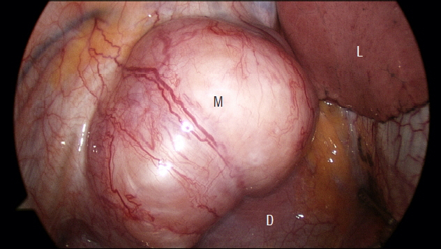
Fig. 1 Intraoperative thoracoscopic finding shows a round mediastinal mass extruding over a diaphragm at T12/L1. M: mass, D: diaphragm, L: lung.
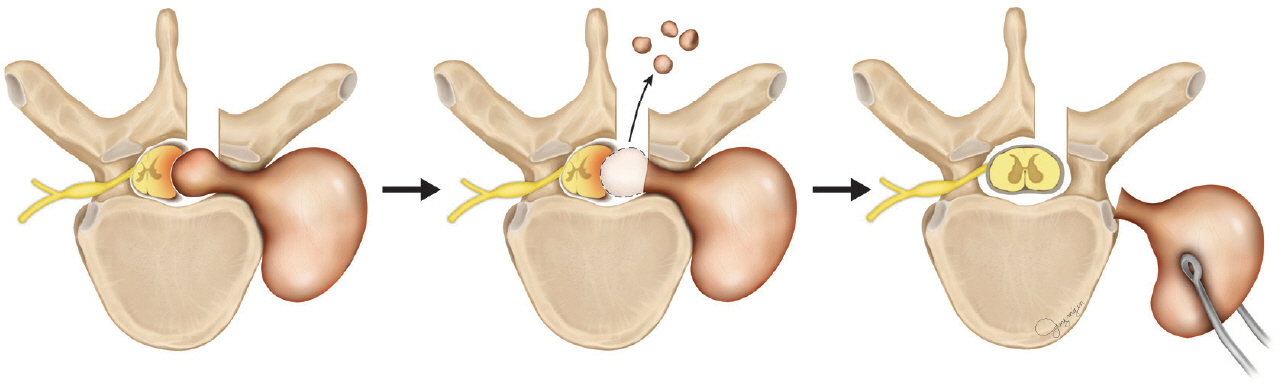
Fig. 2 Schematic illustration of one stage posterior minimal laminectomy and VATS for removal of thoracic dumbbell tumor. VATS: video-assisted thoracoscopic surgery.
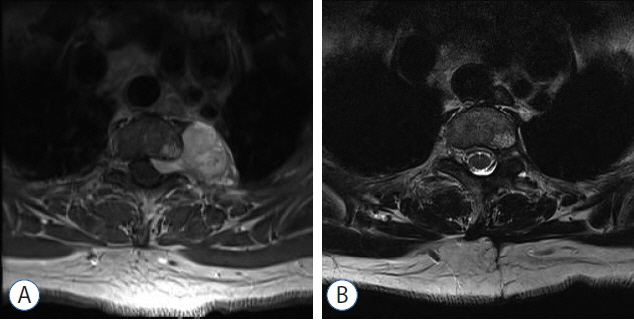
Fig. 3 A: Preoperative gadolinium-enhanced MR image showing a well-enhanced dumbbell shaped tumor with a neuroforaminal extension in the left paravertebral space and posterior mediastinum at T2/3. B: Postoperative T2-weighted MR image shows no remnant tumor and partial laminectomy state reserving lateral facet. MR: magnetic resonance.
Reference
-
References
1. Ando K, Imagama S, Ito Z, Tauchi R, Muramoto A, Matsui H, et al. Removal of thoracic dumbbell tumors through a single-stage posterior approach: its usefulness and limitations. J Orthop Sci. 18:380–387. 2013.
Article2. Ando K, Imagama S, Wakao N, Hirano K, Tauchi R, Muramoto A, et al. Single-stage removal of thoracic dumbbell tumors from a posterior approach only with costotransversectomy. Yonsei Med J. 53:611–617. 2012.
Article3. Hurley JP, McCarthy J, Wood AE. Retrospective analysis of the utility of video-assisted thoracic surgery in 100 consecutive procedures. Eur J Cardiothorac Surg. 8:589–592. 1994.
Article4. Jeon JH, Kang CH, Kim HS, Seong YW, Park IK, Kim YT, et al. Video-assisted thoracoscopic lobectomy in non-small-cell lung cancer patients with chronic obstructive pulmonary disease is associated with lower pulmonary complications than open lobectomy: a propensity score-matched analysis. Eur J Cardiothorac Surg. 45:640–645. 2014.
Article5. Jeong WJ, Choi I, Seong HY, Roh SW. Thoracic extradural cavernous hemangioma mimicking a dumbbell-shaped tumor. J Korean Neurosurg Soc. 58:72–75. 2015.
Article6. Eden K. The dumbbell tumours of the spine. Br J Surg. 28:549–570. 1941.
Article7. Konno S, Yabuki S, Kinoshita T, Kikuchi S. Combined laminectomy and thoracoscopic resection of dumbbell-type thoracic cord tumor. Spine (Phila Pa 1976). 26:E130–E134. 2001.
Article8. Ozawa H, Kokubun S, Aizawa T, Hoshikawa T, Kawahara C. Spinal dumbbell tumors: an analysis of a series of 118 cases. J Neurosurg Spine. 7:587–593. 2007.
Article9. Payer M, Radovanovic I, Jost G. Resection of thoracic dumbbell neurinomas: single postero-lateral approach or combined posterior and transthoracic approach? J Clin Neurosci. 13:690–693. 2006.
Article10. Shiraishi T, Hida S, Isayama T, Yoneda S, Kawahara K, Shirakusa T. A combined thoracoscopic and posterior-spinal approach for “dumbbell” neurofibroma minimizes the anatomical destruction of the vertebrae: report of a case. Surg Today. 32:155–158. 2002.
Article11. Thorat JD, Rajendra T, Thirugnanam A, Ng IH. Single-stage posterior midline approach for dumbbell tumors of the thoracic spine, with intraoperative CT guidance. Surg Neurol Int. 2:31. 2011.
Article12. Vallieres E, Findlay JM, Fraser RE. Combined microneurosurgical and thoracoscopic removal of neurogenic dumbbell tumors. Ann Thorac Surg. 59:469–472. 1995.
Article
- Full Text Links
-
- Actions
-
Cited
- CITED
-
- Close
- Share
-
- Similar articles
-
- Combined Video-Assisted Thoracic Surgery and Posterior Spinal Surgery for the Treatment of Dumbbell Tumor of the First Thoracic Nerve Root
- Video-assisted thoracoscopic lobectomy for lung cancer
- Management of Complications During Video-Assisted Thoracic Surgery Lung Resection and Lymph Node Dissection
- Video-Assisted Thoracic Surgery Pneumonectomy
- Single-Stage Removal of Thoracic Dumbbell Tumors from a Posterior Approach Only with Costotransversectomy
