Use of Three-Dimensional Curved-Multiplanar Reconstruction Images for Sylvian Dissection in Microsurgery of Middle Cerebral Artery Aneurysms
- Affiliations
-
- 1Department of Neurosurgery, Chung-Ang University Hospital, Seoul, Korea. cuttage@cau.ac.kr
- 2Department of Radiology, Chung-Ang University Hospital, Seoul, Korea.
- KMID: 2374212
- DOI: http://doi.org/10.3349/ymj.2017.58.1.241
Abstract
- PURPOSE
The purpose of this study was to introduce a method of using three-dimensional (3D) curved-multiplanar reconstruction (MPR) images for sylvian dissection during microsurgical treatment of middle cerebral artery (MCA) aneurysms.
MATERIALS AND METHODS
Forty-nine patients who had undergone surgery for MCA aneurysms were enrolled. We obtained the 3D curved-MPR images along the sphenoid ridge using OsiriX MDâ„¢ imaging software, compared sylvian dissection time according to several 3D MPR image factors, and investigated the correlations between these images and intraoperative findings.
RESULTS
Utilizing preoperative information of the sylvian fissure (SF) and peri-aneurysmal space on 3D curved-MPR images, we could predict the feasibility of sylvian dissection for a safe surgery. 3D curved-MPR images showed several features: first, perpendicular images to the sylvian surface in the same orientation as the surgeon's view; second, simultaneous visualization of the brain cortex, vessels, and cisternal space; and third, more accurate measurement of various parameters, such as depth of the MCA from the sylvian surface and the location and width of the SFs.
CONCLUSION
In addition to conventional image studies, 3D curved-MPR images seem to provide useful information for Sylvian dissection in the microsurgical treatment of MCA aneurysms.
MeSH Terms
Figure
-
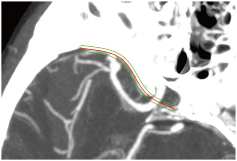
Fig. 1 Curved line along the sphenoid ridge. The line was produced by placing several points along the sphenoid ridge on 3D MPR images. 3D, three-dimensional; MPR, multiplanar reconstruction.
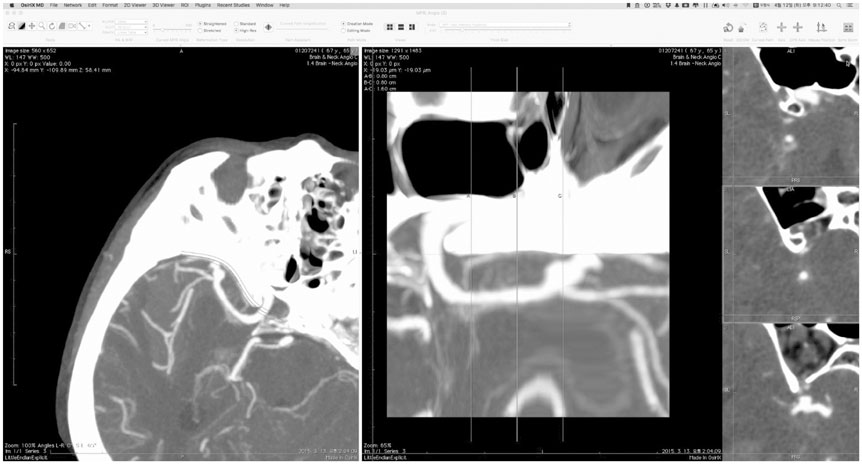
Fig. 2 The working screen. The left panel shows a right MCA aneurysm on the 3D MPR image. The middle panel shows a straightened image of the curved line on the sphenoid ridge, and the right three panels display images perpendicular to the straightened line at three points. MCA, middle cerebral artery; 3D, three-dimensional; MPR, multiplanar reconstruction.
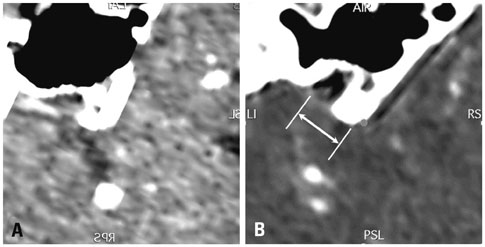
Fig. 3 Location of the sylvian fissure. (A) Central group (<10 mm). (B) Lateral group (≥10 mm). The location of the sylvian fissure (white arrow) is the distance between the anterior clinoid process and the sylvian fissure on the surface. A lateral location of the sylvian fissure means that there is a herniation of the frontal lobe onto the temporal lobe.
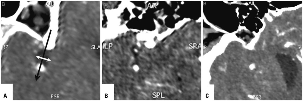
Fig. 4 Width of the sylvian fissure. (A) Wide group (≥3 mm). (B) Narrow group (<3 mm and ≥1 mm). (C) Adhesion group (<1 mm). White arrow is the measurement of the width of the sylvian fissure, which is perpendicular to the direction of dissection corridor (black arrow).

Fig. 5 Depth of the MCA. (A and C) Superficial group [maximal depth (arrow) of the MCA < 10 mm]. (B and D) Deep group [maximal depth (arrow) ≥10 mm]. MCA, middle cerebral artery.
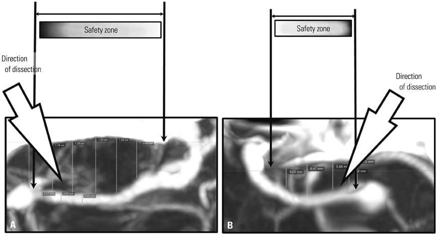
Fig. 6 Safety zone for the arachnoid incision. (A) Long safety zone. (B) Short safety zone. Large arrows reveal the oblique direction of the sylvian dissection. Gradations in the rectangles marked as "safety zone" indicate the degree of risk.

Fig. 7 Intra-operative measurements. Measuring the distance from the anterior clinoid process with a paper ruler.
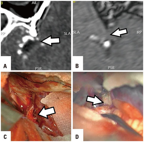
Fig. 8 Cisternal space under the sylvian surface. (A and C) Wide cisternal space (arrows) just under the surface arachnoid membrane. (B and D) Middle cerebral artery and moderate cisternal space under thick cortical adhesion (arrows). This an MR MPR image. MPR, multiplanar reconstruction.
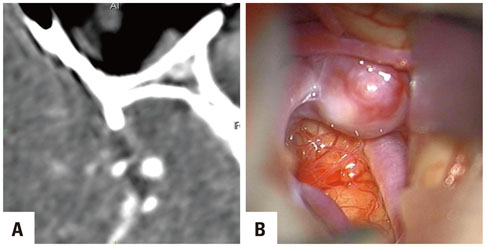
Fig. 9 Location of an aneurysm and MCA branches in the sylvian cistern. (A) MPR image. (B) Intraoperative capture image. Two images provide the following anatomical information: a moderate sylvian cistern, the shallow location of the superior branch, the aneurysm under the temporal lobe, and deep location of the inferior branch. MCA, middle cerebral artery; MPR, multiplanar reconstruction.

Fig. 10 Projection of aneurysmal dome. (A and C) The direction of the aneurysmal dome is opposite to the surface. During sylvian dissection, the secure of aneurysmal neck and branches (arrows) is possible. (B and D) The direction is to sylvian surface, and the dome is located just below superficial sylvian veins on the temporal lobe (arrows).

Fig. 11 Cortical adhesion of aneurysmal dome. (A and C) Aneurysmal dome (arrows) adheres to the frontal lobe. (B and D) Aneurysmal dome (arrows) contacts with the temporal lobe. Temporary clips for safe neck dissection are shown at the proximal and distal site of the aneurysm.
Reference
-
1. Yaşargil MG. Microneurosurgery. New york: Thieme-Stratton;1984.2. Lawton MT. Seven Aneurysms: Tenets and Techniques for Clipping. New York: Thieme;2011.3. Ngando HM, Maslehaty H, Schreiber L, Blaeser K, Scholz M, Petridis AK. Anatomical configuration of the Sylvian fissure and its influence on outcome after pterional approach for microsurgical aneurysm clipping. Surg Neurol Int. 2013; 4:129.
Article4. Huh SK. [Microsurgical anatomy of the middle cerebral artery]. J Korean Neurosurg Soc. 1998; 27:1769–1773.5. Perneczky A, van Lindert E, Müller-Forell W, Fries G. Keyhole concept in neurosurgery. New York: Thieme;1999.6. Harland SP, Hussein A, Gullan RW. Modification of the standard pterional approach for aneurysms of the anterior circle of Willis. Br J Neurosurg. 1996; 10:149–153.
Article7. Hernesniemi J, Ishii K, Niemelä M, Smrcka M, Kivipelto L, Fujiki M, et al. Lateral supraorbital approach as an alternative to the classical pterional approach. Acta Neurochir Suppl. 2005; 94:17–21.
Article8. Lan Q, Gong Z, Kang D, Zhang H, Qian Z, Chen J, et al. Microsurgical experience with keyhole operations on intracranial aneurysms. Surg Neurol. 2006; 66:Suppl 1. S2–S9.
Article9. Mitchell P, Vindlacheruvu RR, Mahmood K, Ashpole RD, Grivas A, Mendelow AD. Supraorbital eyebrow minicraniotomy for anterior circulation aneurysms. Surg Neurol. 2005; 63:47–51.
Article10. Mori K, Osada H, Yamamoto T, Nakao Y, Maeda M. Pterional keyhole approach to middle cerebral artery aneurysms through an outer canthal skin incision. Minim Invasive Neurosurg. 2007; 50:195–201.
Article11. Yamahata H, Tokimura H, Tajitsu K, Tsuchiya M, Taniguchi A, Hirabaru M, et al. Efficacy and safety of the pterional keyhole approach for the treatment of anterior circulation aneurysms. Neurosurg Rev. 2014; 37:629–636.
Article12. Elsharkawy A, Niemelä M, Lehecčka M, Lehto H, Jahromi BR, Goehre F, et al. Focused opening of the sylvian fissure for microsurgical management of MCA aneurysms. Acta Neurochir (Wien). 2014; 156:17–25.
Article13. Ogilvy CS, Crowell RM, Heros RC. Surgical management of middle cerebral artery aneurysms: experience with transsylvian and superior temporal gyrus approaches. Surg Neurol. 1995; 43:15–22.
Article14. Pritz MB, Chandler WF. The transsylvian approach to middle cerebral artery bifurcation/trifurcation aneurysms. Surg Neurol. 1994; 41:217–219.
Article15. Dashti R, Hernesniemi J, Niemelä M, Rinne J, Porras M, Lehecka M, et al. Microneurosurgical management of middle cerebral artery bifurcation aneurysms. Surg Neurol. 2007; 67:441–456.
Article
- Full Text Links
-
- Actions
-
Cited
- CITED
-
- Close
- Share
-
- Similar articles
-
- Superior Temporal Gyrus Approach to Middle Cerebral Artery Aneurysms
- Direction of Middle Cerebral Artery Bifurcation Aneurysms and Surgical Approach
- Two Indices Affecting the Directions of the Sylvian Fissure Dissection in Middle Cerebral Artery Bifurcation Aneurysms
- Surgical Management of Middle Cerebral Artery Aneurysm
- Multiple Aneurysms on the Same Bifurcation Site of the Middle Cerebral Artery
