Performance of and Pressure Elevation Formed by Small-diameter Microtubes Used in Constant-flow Sets
- Affiliations
-
- 1Department of Ophthalmology, Gangneung Asan Hospital, University of Ulsan College of Medicine, Gangneung, Korea.
- 2Department of Mechatronics Engineering, Chungnam National University, Daejeon, Korea.
- 3Malgeunnoon Eye Clinic, Daejeon, Korea.
- 4Department of Ophthalmology, Chungnam National University Hospital, Chungnam National University School of Medicine, Daejeon, Korea. kcs61@cnu.ac.kr
- 5Department of Ophthalmology, Kim's Eye Hospital, Konyang University College of Medicine, Seoul, Korea.
- KMID: 2373978
- DOI: http://doi.org/10.3341/kjo.2016.30.3.225
Abstract
- PURPOSE
We explored the performance of and pressure elevation caused by small-diameter microtubes used to reduce overfiltration.
METHODS
Using a syringe pump-driven constant-flow setting (2 µL/min), pressures were measured for polytetrafluoroethylene (PTFE) microtubes 5 mm in length with inner diameters of 51, 64, and 76 µm and for polyether block amide (PEBAX) microtubes with an inner diameter of 76 µm. Experiments (using microtubes only) were initially performed in air, water, and enucleated pig eyes and were repeated under the same conditions using intraluminal 9/0 nylon stents.
RESULTS
The pressures measured in air in 51-, 64-, and 76-µm-diameter PTFE microtubes differed significantly (22.1, 16.9, and 12.2 mmHg, respectively; p < 0.001), and that of the 76-µm-diameter PEBAX microtube was 15.8 mmHg (p < 0.001 compared to the 12.2 mmHg of the 76-µm-diameter PTFE microtube). The pressures measured in water also differed significantly among the three microtubes at 3.9, 3.0, and 1.4 mmHg, respectively, while that in the PEBAX microtube was 2.6 mmHg (all p < 0.001). Using the intraluminal stent, the pressure in water of the three different PTFE microtubes increased to 22.6, 18.0, and 4.1 mmHg, respectively, and that in the PEBAX microtube increased to 10.5 mmHg (all p < 0.001). Similar trends were evident when measurements were performed in pig eyes.
CONCLUSIONS
Although microtubes of smaller diameter experienced higher pressure in air, reduction of the inner diameter to 51 µm did not adequately increase the pressure attained in water or pig eyes. Insertion of an intraluminal stent effectively elevated the latter pressures. PEBAX microtubes created higher pressures than did PTFE microtubes.
Keyword
MeSH Terms
Figure
-

Fig. 1 Light micrographs of the cut ends, and average internal diameters of the microtubes and 9/0 nylon thread. (A) 51-µm polytetrafluoroethylene (PTFE) tube. (B) 64-µm PTFE tube. (C) 76-µm PTFE tube. (D) 76-µm polyether block amide tube. (E) 9/0 nylon. (F) Tube with a 20-mm length of intraluminal 9/0 nylon; one end of the thread was inserted into the tube 6 mm from the distal end.

Fig. 2 Experimental setup for fluid flow in air. The setup consisted of a perfusion pump with a syringe, pressure transducer, pressure detector, silicone chamber, and microtube.
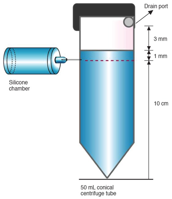
Fig. 3 Experimental setup for fluid flow in water. All settings were identical to those in Fig. 2, except that the tip of the microtube was connected to a tube filled with distilled water.
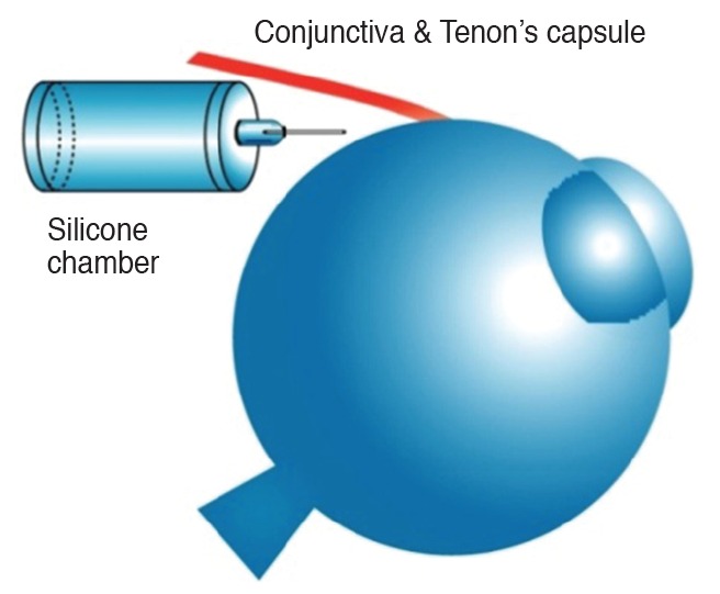
Fig. 4 Experimental setup for fluid flow in an enucleated pig's eye. All settings are identical to those in Fig. 2, except the tip of the microtube was positioned on the sclera beneath the conjunctiva and Tenon's capsule.

Fig. 5 Cumulative pressure profiles for microtubes of different diameters and materials. (A) Microtubes without a stent in air. (B) Microtubes with an intraluminal 9/0 nylon stent in air. (C) Microtubes without a stent, connected to a water chamber. (D) Microtubes with an intraluminal 9/0 nylon stent, connected to a water chamber. (E) Microtubes without a stent, connected to a pig eye. (F) Microtubes with an intraluminal 9/0 nylon stent, connected to a pig eye. PTFE = polytetrafluoroethylene; PEBAX = polyether block amide.
Cited by 1 articles
-
Fluid Dynamics of Small Diameter Tubes Used in Membrane-tube Type Glaucoma Shunt Devices
Jong Chul Han, Young Hoon Hwang, Byung Heon Ahn
Korean J Ophthalmol. 2019;33(4):371-378. doi: 10.3341/kjo.2019.0027.
Reference
-
1. Lieberman MF, Ewing RH. Drainage implant surgery for refractory glaucoma. Int Ophthalmol Clin. 1990; 30:198–208. PMID: 2199389.
Article2. Heuer DK, Lloyd MA, Abrams DA, et al. Which is better? One or two? A randomized clinical trial of single-plate versus double-plate Molteno implantation for glaucomas in aphakia and pseudophakia. Ophthalmology. 1992; 99:1512–1519. PMID: 1454316.3. Drake M. Complications of glaucoma filtration surgery. Int Ophthalmol Clin. 1992; 32:115–130. PMID: 1399343.
Article4. Melamed S, Cahane M, Gutman I, Blumenthal M. Postoperative complications after Molteno implant surgery. Am J Ophthalmol. 1991; 111:319–322. PMID: 2000901.
Article5. Sarkisian SR Jr. Tube shunt complications and their prevention. Curr Opin Ophthalmol. 2009; 20:126–130. PMID: 19240545.
Article6. Krupin T, Podos SM, Becker B, Newkirk JB. Valve implants in filtering surgery. Am J Ophthalmol. 1976; 81:232–235. PMID: 814819.
Article7. Egbert PR, Lieberman MF. Internal suture occlusion of the Molteno glaucoma implant for the prevention of postoperative hypotony. Ophthalmic Surg. 1989; 20:53–56. PMID: 2648233.
Article8. Price FW Jr, Whitson WE. Polypropylene ligatures as a means of controlling intraocular pressure with Molteno implants. Ophthalmic Surg. 1989; 20:781–783. PMID: 2616124.
Article9. Sherwood MB, Smith MF. Prevention of early hypotony associated with Molteno implants by a new occluding stent technique. Ophthalmology. 1993; 100:85–90. PMID: 8433833.
Article10. Hoare Nairne JE, Sherwood D, Jacob JS, Rich WJ. Single stage insertion of the Molteno tube for glaucoma and modifications to reduce postoperative hypotony. Br J Ophthalmol. 1988; 72:846–851. PMID: 3207660.
Article11. Molteno AC, Polkinghorne PJ, Bowbyes JA. The vicryl tie technique for inserting a draining implant in the treatment of secondary glaucoma. Aust N Z J Ophthalmol. 1986; 14:343–354. PMID: 3814422.
Article12. Smith MF, Doyle JW, Ticrney JW Jr. A comparison of glaucoma drainage implant tube coverage. J Glaucoma. 2002; 11:143–147. PMID: 11912362.
Article13. Heuer DK, Budenz D, Coleman A. Aqueous shunt tube erosion. J Glaucoma. 2001; 10:493–496. PMID: 11740221.
Article14. Prata JA Jr, Minckler DS, Mermoud A, Baerveldt G. Effects of intraoperative mitomycin-C on the function of Baerveldt glaucoma drainage implants in rabbits. J Glaucoma. 1996; 5:29–38. PMID: 8795731.
Article15. Lim KS, Wells AP, Khaw PT. Needle perforations of Molteno tubes. J Glaucoma. 2002; 11:434–438. PMID: 12362085.
Article16. Prata JA Jr, Mermoud A, LaBree L, Minckler DS. In vitro and in vivo flow characteristics of glaucoma drainage implants. Ophthalmology. 1995; 102:894–904. PMID: 7777296.
Article17. McEWEN WK. Application of Poiseuille's law to aqueous outflow. AMA Arch Ophthalmol. 1958; 60:290–294. PMID: 13558800.
Article18. Minckler DS, Heuer DK, Hasty B, et al. Clinical experience with the single-plate Molteno implant in complicated glaucomas. Ophthalmology. 1988; 95:1181–1188. PMID: 3211496.
Article19. Molteno AC, Van Biljon G, Ancker E. Two-stage insertion of glaucoma drainage implants. Trans Ophthalmol Soc N Z. 1979; 31:17–26. PMID: 293078.20. Francis BA, Singh K, Lin SC, et al. Novel glaucoma procedures: a report by the American Academy of Ophthalmology. Ophthalmology. 2011; 118:1466–1480. PMID: 21724045.21. White FM, editor. Fluid mechanics. 6th ed. New York: Mc-Graw-Hill;2008. p. 31–34.22. Hetsroni G, Mosyak A, Pogrebnyak E, Yarin LP. Fluid flow in micro-channels. Int J Heat Mass Transf. 2005; 48:1982–1998.
Article23. Li ZX. Experimental study on flow characteristics of liquid in circular microtubes. Microscale Thermophys Eng. 2003; 7:253–265.
Article24. Civan MM, Benos DJ, Simon SA, editors. The eye's aqueous humor. 2nd ed. San Diego: Academic Press;2008. p. 174–175.
- Full Text Links
-
- Actions
-
Cited
- CITED
-
- Close
- Share
-
- Similar articles
-
- Changes in the Rate of Flow by Quantitative Analysis of Aqueous Humor after Argon Laser Trabeculoplasty in Rabbits
- Changes of the Vital Sign, Cerebral Perfusion Pressure and Intracranial Pressure in Variable Degree of Head Elevation
- Hemodynamic effects of the geometric dimensions of graft vessels in coronary artery bypass graft models
- Comparison of the Clinical Effects of the Different Ventilatory Care Strategies in the Neonates with Acute Respiratory Failure: High Flow Rate - Constant Flow Rate
- Difference in Patient's Work of Breathing Between Pressure-Controlled Ventilation with Deccelerating Flow and Volume-Controlled Ventilation with Constant Flow during Assited Ventilation
