Korean J Ophthalmol.
2016 Jun;30(3):163-171. 10.3341/kjo.2016.30.3.163.
Angiogenin for the Diagnosis and Grading of Dry Eye Syndrome
- Affiliations
-
- 1Department of Ophthalmology, Chung-Ang University Hospital, Chung-Ang University College of Medicine, Seoul, Korea. jck50ey@daum.net
- KMID: 2373971
- DOI: http://doi.org/10.3341/kjo.2016.30.3.163
Abstract
- PURPOSE
To investigate the properties of angiogenin (ANG) as a potential tool for the diagnosis and grading of dry eye syndrome (DES) by analyzing tear protein profiles.
METHODS
Tear samples were collected with capillary tubes from 52 DES patients and 29 normal individuals as controls. Tear protein profiles were analyzed with an immunodot blot assay as a screening test. To confirm that the tear ANG levels were in inverse proportion to the disease severity grade, the ANG and lactoferrin (LF) tear contents of normal controls and DES patients were compared in an enzyme-linked immunosorbent assay.
RESULTS
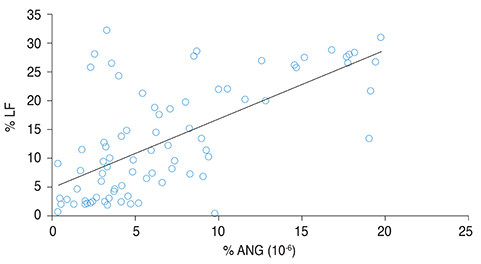
In the immunodot blot assay, the ANG area was lower in patients with grades 3 and 4 DES than in normal controls. The areas of basic fibroblast growth factor, transforming growth factor β2, and interleukin 10 were significantly greater than those of normal controls only in grade 4 DES patients, but these proteins were not linearly correlated with dry eye severity. Upon enzyme-linked immunosorbent assay analysis, the mean concentrations of ANG and LF decreased significantly as dry eye severity increased, except between grades 1 and 2. In addition, the ratios of ANG and LF to total tear proteins were correlated significantly with DES severity.
CONCLUSIONS
ANG level was significantly lower in DES patients than in normal controls, and was significantly correlated with the worsening severity of DES, except between grades 1 and 2, as was LF. Therefore, ANG may be a useful measure of DES severity through proteomic analysis.
Keyword
MeSH Terms
-
Adult
Aged
Angiogenesis Inducing Agents/pharmacology
Dry Eye Syndromes/*diagnosis/metabolism
Enzyme-Linked Immunosorbent Assay
Female
Follow-Up Studies
Humans
Immunoblotting
Male
Middle Aged
Proteomics/methods
Ribonuclease, Pancreatic/*pharmacology
Severity of Illness Index
Tears/chemistry
Young Adult
Angiogenesis Inducing Agents
Ribonuclease, Pancreatic
Figure
Reference
-
1. Javadi MA, Feizi S. Dry eye syndrome. J Ophthalmic Vis Res. 2011; 6:192–198.2. Bhavsar AS, Bhavsar SG, Jain SM. A review on recent advances in dry eye: pathogenesis and management. Oman J Ophthalmol. 2011; 4:50–56.3. Stern ME, Gao J, Siemasko KF, et al. The role of the lacrimal functional unit in the pathophysiology of dry eye. Exp Eye Res. 2004; 78:409–416.4. Pflugfelder SC. Anti-inf lammatory therapy of dry eye. Ocul Surf. 2003; 1:31–36.5. Solomon A, Dursun D, Liu Z, et al. Pro- and anti-inflammatory forms of interleukin-1 in the tear fluid and conjunctiva of patients with dry-eye disease. Invest Ophthalmol Vis Sci. 2001; 42:2283–2292.6. Rolando M, Barabino S, Mingari C, et al. Distribution of conjunctival HLA-DR expression and the pathogenesis of damage in early dry eyes. Cornea. 2005; 24:951–954.7. Farris RL. Tear osmolarity: a new gold standard? Adv Exp Med Biol. 1994; 350:495–503.8. Gao J, Schwalb TA, Addeo JV, et al. The role of apoptosis in the pathogenesis of canine keratoconjunctivitis sicca: the effect of topical Cyclosporin A therapy. Cornea. 1998; 17:654–663.9. de Souza GA, Godoy LM, Mann M. Identification of 491 proteins in the tear fluid proteome reveals a large number of proteases and protease inhibitors. Genome Biol. 2006; 7:R72.10. Green-Church KB, Nichols KK, Kleinholz NM, et al. Investigation of the human tear film proteome using multiple proteomic approaches. Mol Vis. 2008; 14:456–470.11. Versura P, Nanni P, Bavelloni A, et al. Tear proteomics in evaporative dry eye disease. Eye (Lond). 2010; 24:1396–1402.12. Weremowicz S, Fox EA, Morton CC, Vallee BL. Localization of the human angiogenin gene to chromosome band 14q11, proximal to the T cell receptor alpha/delta locus. Am J Hum Genet. 1990; 47:973–981.13. Gao X, Xu Z. Mechanisms of action of angiogenin. Acta Biochim Biophys Sin (Shanghai). 2008; 40:619–624.14. Lee SH, Kim KW, Min KM, et al. Angiogenin reduces immune inflammation via inhibition of TANK-binding kinase 1 expression in human corneal fibroblast cells. Mediators Inflamm. 2014; 2014:861435.15. Sack RA, Conradi L, Krumholz D, et al. Membrane array characterization of 80 chemokines, cytokines, and growth factors in open- and closed-eye tears: angiogenin and other defense system constituents. Invest Ophthalmol Vis Sci. 2005; 46:1228–1238.16. The definition and classification of dry eye disease: report of the Definition and Classification Subcommittee of the International Dry Eye WorkShop (2007). Ocul Surf. 2007; 5:75–92.17. McMonnies CW. Key questions in a dry eye history. J Am Optom Assoc. 1986; 57:512–517.18. Nichols KK, Nichols JJ, Mitchell GL. The lack of association between signs and symptoms in patients with dry eye disease. Cornea. 2004; 23:762–770.19. Aebersold R, Mann M. Mass spectrometry-based proteomics. Nature. 2003; 422:198–207.20. Oh JY, Kim MK, Choi HJ, et al. Investigating the relationship between serum interleukin-17 levels and systemic immune-mediated disease in patients with dry eye syndrome. Korean J Ophthalmol. 2011; 25:73–76.21. Zittermann SI, Issekutz AC. Basic fibroblast growth factor (bFGF, FGF-2) potentiates leukocyte recruitment to inflammation by enhancing endothelial adhesion molecule expression. Am J Pathol. 2006; 168:835–846.22. Pflugfelder SC, Stern ME. Symposium Participants. Immunoregulation on the ocular surface: 2nd Cullen Symposium. Ocul Surf. 2009; 7:67–77.23. Danjo Y, Lee M, Horimoto K, Hamano T. Ocular surface damage and tear lactoferrin in dry eye syndrome. Acta Ophthalmol (Copenh). 1994; 72:433–437.24. Tsai PS, Evans JE, Green KM, et al. Proteomic analysis of human meibomian gland secretions. Br J Ophthalmol. 2006; 90:372–377.25. Conneely OM. Antiinflammatory activities of lactoferrin. J Am Coll Nutr. 2001; 20:5 Suppl. 389S–395S.26. Brock JH. Lactoferrin: 50 years on. Biochem Cell Biol. 2012; 90:245–251.27. Dogru M, Matsumoto Y, Yamamoto Y, et al. Lactoferrin in Sjogren's syndrome. Ophthalmology. 2007; 114:2366–2367.28. Murata M, Wakabayashi H, Yamauchi K, Abe F. Identification of milk proteins enhancing the antimicrobial activity of lactoferrin and lactoferricin. J Dairy Sci. 2013; 96:4891–4898.29. Schmaldienst S, Oberpichler A, Tschesche H, Horl WH. Angiogenin: a novel inhibitor of neutrophil lactoferrin release during extracorporeal circulation. Kidney Blood Press Res. 2003; 26:107–112.30. Skeie JM, Zeng S, Faidley EA, Mullins RF. Angiogenin in age-related macular degeneration. Mol Vis. 2011; 17:576–582.31. Marek N, Raczynska K, Siebert J, et al. Decreased angiogenin concentration in vitreous and serum in proliferative diabetic retinopathy. Microvasc Res. 2011; 82:1–5.32. Cho P, Brown B, Chan I, et al. Reliability of the tear breakup time technique of assessing tear stability and the locations of the tear break-up in Hong Kong Chinese. Optom Vis Sci. 1992; 69:879–885.33. Abusharha AA, Pearce EI. The effect of low humidity on the human tear film. Cornea. 2013; 32:429–434.
- Full Text Links
- Actions
-
Cited
- CITED
-
- Close
- Share
- Similar articles
-
- The significance of tear film break-up time in the diagnosis of dry eye syndrome
- Diagnostic Criteria for Sjoren's Syndrome
- Dry Eye Assessment of Patients Undergoing Endoscopic Dacryocystorhinostomy for Nasolacrimal Duct Obstruction Combined with Dry Eye Syndrome
- Bacterial Conjuntival Flora in Dry Eye Patients
- Inhibitory Effect of Rapamycin on Corneal Neovascularization induced by Angiogenin in Rabbits


