The Results of the First Ray Forefoot Osteotomy Using Low Profile Wedge Plate without a Bone Grafting for Pes Planus Correction
- Affiliations
-
- 1W Institute for Foot and Ankle Disease and Trauma, W Hospital, Daegu, Korea.
- 2Department of Orthopedic Surgery, Ilsan Paik Hospital, Inje University College of Medicine, Goyang, Korea. sjs0506@paik.ac.kr
- KMID: 2371967
- DOI: http://doi.org/10.14193/jkfas.2017.21.1.7
Abstract
- PURPOSE
We retrospectively analyzed the radiographic and clinical results after the first ray of forefoot osteotomy using low profile wedge plate without additional cancellous bone grafting for pes planus correction.
MATERIALS AND METHODS
Twenty-four patients were enrolled in this study. Medial cuneiform opening wedge osteotomy was performed in 12 patients (Cotton osteotomy, group C) and first metatarsal base osteotomy was performed in 12 patients (group MT).
RESULTS
On average, the wedge size was 5.61 mm (5~6 mm). The mean time to radiographic union was 3.18 and 3.27 months in groups C and MT, respectively. Postoperative talonavicular coverage angle, talo-first metatarsal angle (anteroposterior), talo-first metatarsal angle (lateral), talo-calcaneal angle (lateral), medial cuneiform height, and American orthopaedic foot, as well as ankle society midfoot scale were significantly improved in both groups. Nonunion, delayed union or fixation failure was not presented in our series.
CONCLUSION
We have shown that low profile wedge plate was effective in the case of first ray forefoot osteotomy for pes planus correction without any additional cancellous bone grafting.
Keyword
MeSH Terms
Figure
-

Figure 1. Various types of low profile mini opening wedge plate used for pes planus deformity correction (Arthrex Inc., Naples, FL, USA).

Figure 2. Preoperative weightbearing foot anteroposterior (A) and lateral (B) radiographs of a 37-year-old male patient with pes planus. (C, D) Immediate postoperative radiographs shows the correction of radiographic parameters. Note that the opening wedge gap in the medial cuneiform bone was remained empty without additional cancellous bone graft. (E, F) Radiographs at the final follow-up (1.5 years) show a stable maintenance with a radiographic union.
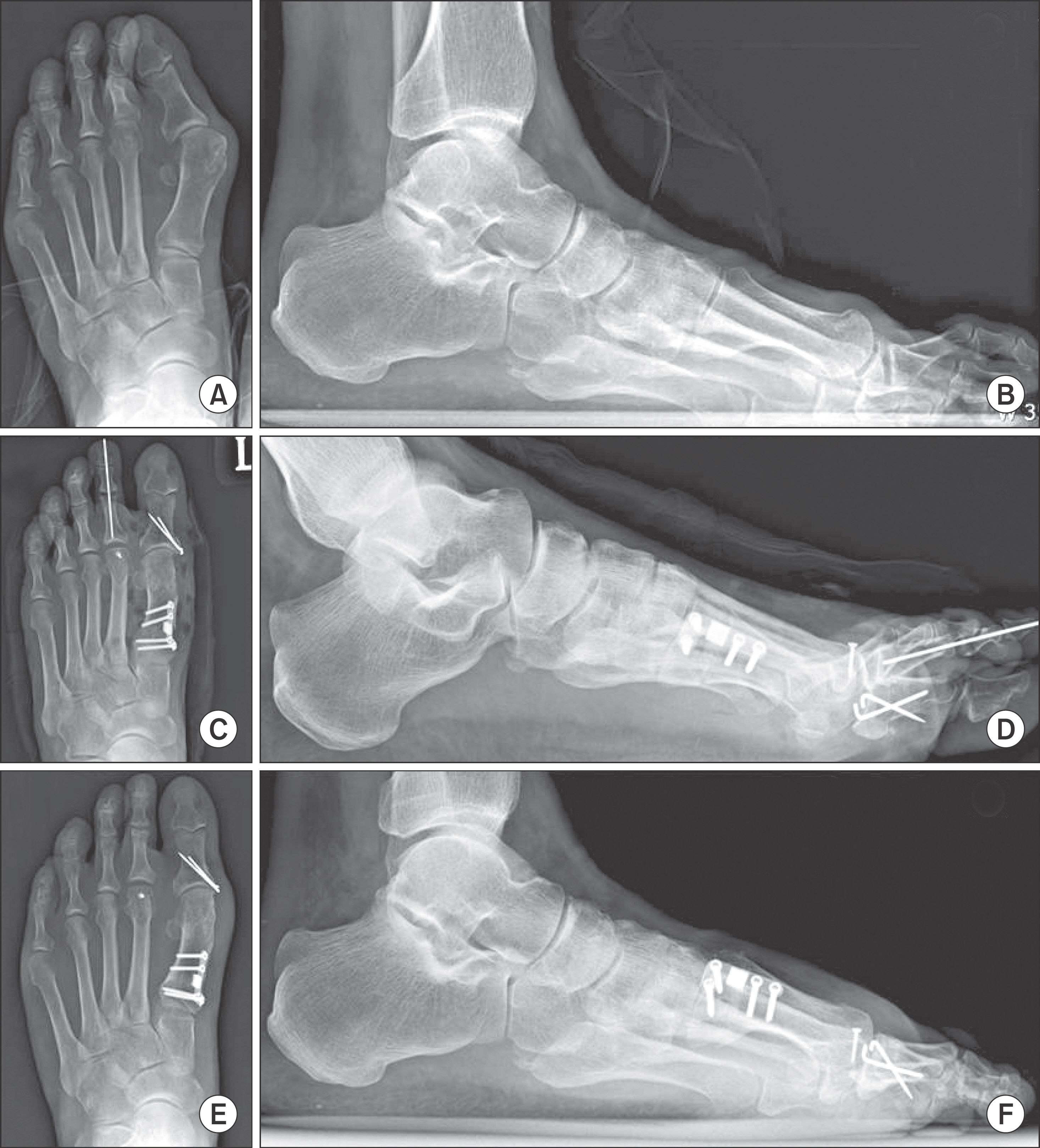
Figure 3. Preoperative weightbearing foot anteroposterior (A) and lateral (B) radiographs of a 47-year-old female patient with pes planus and hallux valgus deformity. (C, D) Immediate postoperative radiographs shows the correction of radiographic parameters. Note that the opening wedge gap in the 1st metatarsal bone was remained empty without additional cancellous bone graft. (E, F) Radiographs at the final follow-up (1 year) show a stable maintenance with a radiographic union.
Reference
-
1.Cohen BE., Ogden F. Medial column procedures in the acquired flatfoot deformity. Foot Ankle Clin. 2007. 12:287–99.
Article2.Mosier-LaClair S., Pomeroy G., Manoli A 2nd. Operative treatment of the difficult stage 2 adult acquired flatfoot deformity. Foot Ankle Clin. 2001. 6:95–119.
Article3.Sizensky JA., Marks RM. Medial-sided bony procedures: why, what, and how? Foot Ankle Clin. 2003. 8:539–62.
Article4.Cotton FJ. Foot statics and surgery. N Engl J Med. 1936. 214:353–62.
Article5.Han JH., Kim HJ., Song JG., Yang JH., Bhandare NN., Fernandez AR, et al. Is bone grafting necessary in opening wedge high tibial osteotomy? A meta-analysis of radiological outcomes. Knee Surg Relat Res. 2015. 27:207–20.
Article6.El-Assal MA., Khalifa YE., Abdel-Hamid MM., Said HG., Bakr HM. Opening-wedge high tibial osteotomy without bone graft. Knee Surg Sports Traumatol Arthrosc. 2010. 18:961–6.
Article7.Zorzi AR., da Silva HG., Muszkat C., Marques LC., Cliquet A Jr., de Miranda JB. Opening-wedge high tibial osteotomy with and without bone graft. Artif Organs. 2011. 35:301–7.
Article8.Kitaoka HB., Alexander IJ., Adelaar RS., Nunley JA., Myerson MS., Sanders M. Clinical rating systems for the ankle-hindfoot, midfoot, hallux, and lesser toes. Foot Ankle Int. 1994. 15:349–53.
Article9.Hirose CB., Johnson JE. Plantarflexion opening wedge medial cuneiform osteotomy for correction of fixed forefoot varus associated with flatfoot deformity. Foot Ankle Int. 2004. 25:568–74.
Article10.Castaneda D., Thordarson DB., Charlton TP. Radiographic assessment of medial cuneiform opening wedge osteotomy for flatfoot correction. Foot Ankle Int. 2012. 33:498–500.
Article11.Tankson CJ. The Cotton osteotomy: indications and techniques. Foot Ankle Clin. 2007. 12:309–15.
Article12.Yarmel D., Mote G., Treaster A. The cotton osteotomy: a technical guide. J Foot Ankle Surg. 2009. 48:506–12.
Article13.Van Gestel L., Van Bouwel S., Somville J. Surgical treatment of the adult acquired flexible flatfoot. Acta Orthop Belg. 2015. 81:172–83.14.Needleman RL. A surgical approach for flexible flatfeet in adults including a subtalar arthroereisis with the MBA sinus tarsi im-plant. Foot Ankle Int. 2006. 27:9–18.
Article15.League AC., Parks BG., Schon LC. Radiographic and pedobaro-graphic comparison of femoral head allograft versus block plate with dorsal opening wedge medial cuneiform osteotomy: a biomechanical study. Foot Ankle Int. 2008. 29:922–6.
Article
- Full Text Links
-
- Actions
-
Cited
- CITED
-
- Close
- Share
-
- Similar articles
-
- The Clinical Results of the Proximal Opening Wedge Osteotomy Using a Low Profile Plate in Hallux Valgus: Comparison with Proximal Chevron Osteotomy Fixed with K-wires
- Back out of Locking Pin with Hinge Fracture after High Tibial Osteotomy
- Severe Genu Recurvatum after a Closing-wedge High Tibial Osteotomy: A Case Report
- First Metatarsal Proximal Opening Wedge Osteotomy for Correction of Hallux Valgus Deformity: Comparison of Straight versus Oblique Osteotomy
- Can medial stability be preserved after open wedge high tibial osteotomy?
