The Results of Medial Horizontal Suture Fixation of Akin Osteotomy in Hallux Valgus
- Affiliations
-
- 1Orthopaedic Surgery Department, Daejeon Woori Hospital, Daejeon, Korea. yuneyp@naver.com
- KMID: 2371966
- DOI: http://doi.org/10.14193/jkfas.2017.21.1.1
Abstract
- PURPOSE
The purpose of this study was to analyze the clinical results of medial horizontal suture fixation of Akin osteotomy in hallux valgus and present its advantages.
MATERIALS AND METHODS
This study was based on 48 cases of 35 patients with Akin osteotomy, who underwent surgery of hallux valgus between December 2014 and July 2015, and with at least 12 months of follow-up. The mean age of patients was 46.9 years (range, 16~71 years). The mean follow-up duration was 15.9 months (range, 12~18 months). Clinical evaluations included pain visual analogue scale (VAS) score, American Orthopaedic Foot and Ankle Society (AOFAS hallux metatarsophalangeal interphalangeal scale) score, and satisfaction score. Weightbearing anteroposterior radiographs were taken to measure the distal articular set angle (DASA) of the hallux. Radiographic bone union at 6 months follow-up was regarded as a success, while a loss of reduction and nonunion was regarded as a failure.
RESULTS
The mean pre- and postoperative pain VAS scores were 4.27 and 1.67, respectively (p<0.05). The mean AOFAS score improved from 59.7 to 80.5 (p<0.05). The DASA was improved from 8.15 to -2.57 (p<0.05). There was no case of skin irritation, cortical breakage, inflammation from the knot, and infection. All patients showed union without fixation failure.
CONCLUSION
The clinical and radiological evaluations in this study demonstrate reliable results without complication. The medial horizontal suture fixation of the Akin osteotomy was effective, and the advantage of this procedure was unnecessity of the material removal, preservation of the joint, and no skin irritation.
Keyword
MeSH Terms
Figure
-
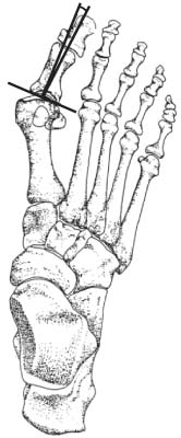
Figure 1 Distal articular set angle.

Figure 2 Osteotomy is performed on the proximal phalanx 5 to 6 mm distal to the first metatarsophalangeal joint. The size of medial closing wedge osteotomy was determined by remaining valgus angle. (A) Anterior view. (B) Lateral view.
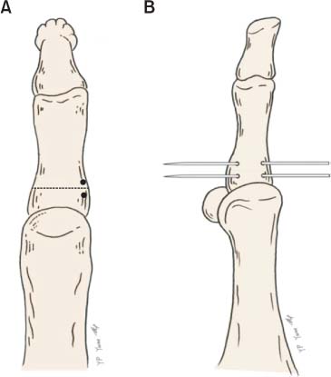
Figure 3 Two holes are placed horizontally on either side of osteotomy using 1.6 mm Kirschner wire. (A) Anterior view. (B) Lateral view.
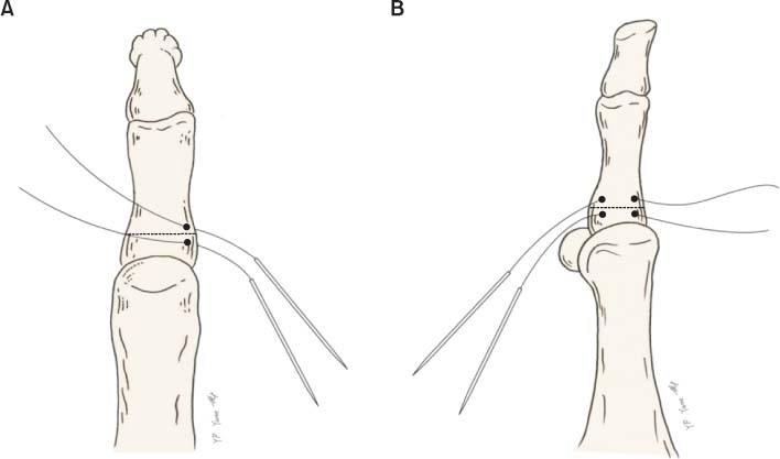
Figure 4 No. 2 Ethibond sutures (Ethicon, USA) on straight needle are passed into both side holes. (A) Anterior view. (B) Lateral view.

Figure 5 Osteotomy site was closed and tied with Ethibond suture (Ethicon, USA). (A) Anterior view. (B) Lateral view.
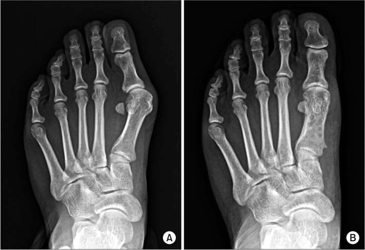
Figure 6 (A) Preoperative weightbearing anteroposterior radiograph of 47-year-old woman shows hallux valgus in the left foot. (B) Radiograph at postoperative 18 months shows bone union.
Reference
-
1. Akin OF. The treatment of hallux valgus: a new operative procedure and its results. Med Sentinel. 1925; 33:678–679.2. Yune YP, Kim S. Medial horizontal suture fixation of the Akin osteotomy: a technical report. J Korean Foot Ankle Soc. 2015; 19:197–200.
Article3. Mau C, Lauber HJ. Die operative Behandlung des Hallux valgus (Nachuntersuchungen). Deutsche Zeitschrift für Chirurgie. 1926; 197:361–377.
Article4. Balding MG, Sorto LA Jr. Distal articular set angle. Etiology and x-ray evaluation. J Am Podiatr Med Assoc. 1985; 75:648–652.
Article5. Springer KR. The role of the Akin osteotomy in the surgical management of hallux abducto valgus. Clin Podiatr Med Surg. 1989; 6:115–131.6. Gerbert J, Spector E, Clark J. Osteotomy procedures on the proximal phalanx for correction of a hallux deformity. J Am Podiatry Assoc. 1974; 64:617–629.
Article7. Colloff B, Weitz EM. Proximal phalangeal osteotomy in hallux valgus. Clin Orthop Relat Res. 1967; 54:105–113.
Article8. Coughlin MJ. Hallux valgus. J Bone Joint Surg Am. 1996; 78:932–966.9. Levitsky DR, DiGilio J, Kander R, Rubin B. Rigid compression screw fixation of first proximal phalanx osteotomy for hallux abducto valgus. J Foot Surg. 1982; 21:65–69.10. Giannestras NJ. Foot disorders: medical and surgical management. 2nd ed. Philadelphia: Lea & Febiger;1973.11. Tóth K, Kellermann P, Wellinger K. Fixation of Akin osteotomy for hallux abductus with absorbable suture. Arch Orthop Trauma Surg. 2010; 130:1257–1261.
Article12. Chacon Y, Fallat LM, Dau N, Bir C. Biomechanical comparison of internal fixation techniques for the Akin osteotomy of the proximal phalanx. J Foot Ankle Surg. 2012; 51:561–565.
Article13. Boberg J. Surgical procedures of the hallux. In : McGlamary ED, Banks AS, Downey M, editors. The comprehensive textbook of foot and ankle surgery. 2nd ed. Philadelphia: JB Lippincott;1992. p. 533–534.14. Schlefman BS. Akin osteotomy with horizontal interosseous wire-loop fixation. J Am Podiatr Med Assoc. 1999; 89:194–198.
Article15. Song MH, Kim BH, Ahn SJ, Yoo SH, Lee DJ. Fixation with absorbable suture material in Akin osteotomy. J Korean Foot Ankle Soc. 2011; 15:149–152.16. Ahn SJ, Kim BH, Song MH, Kang SW, Oh KT, Yoo SH. Transarticular fixation of Akin osteotomy on patients with hallux valgus after resection of medial protrusion of base of proximal phalanx. J Korean Foot Ankle Soc. 2013; 17:220–224.17. Moon GH, Ahn GY, Lee YH, Nam IH, Lee JI. The effect of derotational closing wedge Akin osteotomy for the treatment of hallux valgus with the pronation of great toe. Korean Foot Ankle Soc. 2008; 12:14–19.18. Young KW, Lee KT, Kim JY, Cha SD, Kim ES. Fixation with suture material in Akin osteotomy. J Korean Foot Ankle Soc. 2004; 8:138–141.
- Full Text Links
-
- Actions
-
Cited
- CITED
-
- Close
- Share
-
- Similar articles
-
- Medial Horizontal Suture Fixation of the Akin Osteotomy: A Technical Report
- Fixation with Absorbable Suture Material in Akin Osteotomy
- Transarticular Fixation of Akin Osteotomy on Patients with Hallux Valgus after Resection of Medial Protrusion of Base of Proximal Phalanx
- The Effect of Derotational Closing Wedge Akin Osteotomy for the Treatment of Hallux Valgus with the Pronation of Great Toe
- Fixation with Suture Material in Akin Osteotomy
