Surgical Techniques for Percutaneous Intramedullary Fixation with Steinmann Pins for Clavicle Shaft Fractures
- Affiliations
-
- 1Department of Orthopedic Surgery, Sahmyook Medical Center, Seoul, Korea. crash1401@naver.com
- KMID: 2369566
- DOI: http://doi.org/10.4055/jkoa.2017.52.1.7
Abstract
- PURPOSE
To report the clinical results from surgical treatment for clavicle shaft fracture by percutaneous intramedullary fixation with Steinmann pins.
MATERIALS AND METHODS
Between January 2004 and June 2014, the medical records of 135 patients who underwent percutaneous intramedullary fixation with Steinmann pins were reviewed. The mean follow-up periods were 15 months. The functional results were evaluated with The Disabilities of the Arm, Shoulder and Hand (DASH) score and Constant score. The clinical results were evaluated with the shortened length of the clavicle, length of surgical wound, operation time and Kang's criteria.
RESULTS
The mean bone union period was 11.6 weeks (8-16 weeks). The mean DASH score was 11.8. The mean Constant score was 91.2. The mean shortened length of the clavicle was less than 20 mm. The mean length of surgical wound was 1.2 cm (0.7-1.5 cm). The mean operation time was 18 minutes (10-35 minutes). Using Kang's criteria, 131 out of 135 patients (97.0%) showed good results. Complications included were 3 pin migrations and 2 non-unions.
CONCLUSION
Percutaneous intramedullary fixation with Steinmann pins showed good results for treating clavicle shaft fracture.
Figure
-
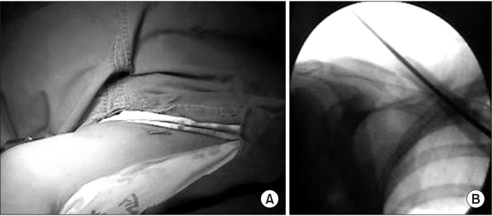
Figure 1 Photograph (A) and radiograph (B) showing percutaneous insertion of a Steinmann pin into the intramedullary canal under fluoroscopic guidance from the fracture side to the medial end.

Figure 2 Photograph (A) and radiograph (B) showing a reduction of the clavicle fracture with a towel clip.

Figure 3 Photograph (A) and radiograph (B) showing retrograde reinsertion of a Steinmann pin under fluoroscopic guidance after fracture site reduction.
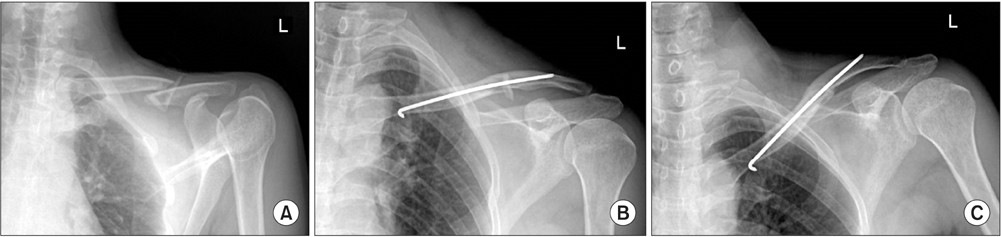
Figure 4 (A) Picture shows the displaced clavicle shaft fracture as Robinson classification type 2B1. (B) Immediate postoperative radiograph shows a well-reducted clavicle with percutaneous Steinmann pin fixation. (C) Radiograph from postoperative 12 weeks shows bone union.
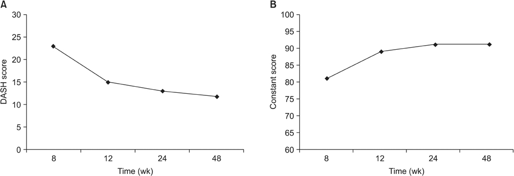
Figure 5 In the functional assessment, postoperative Disabilities of the Arm, Shoulder and Hand (DASH) score (A) and Constant score (B) change.
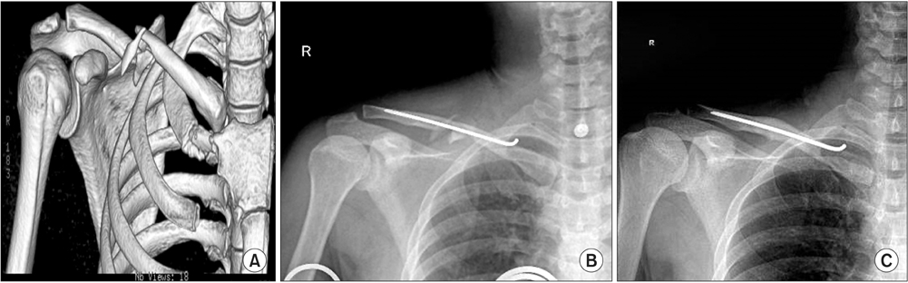
Figure 6 (A) Preoperative three-dimensional computed tomographic image showing a displaced clavicle shaft fracture with a double-butterfly fragment classified as Robinson type 2B2. (B) Immediate postoperative radiograph showing a well-reducted clavicle with double butterfly fragments with Steinmann pin fixation. (C) Radiograph from postoperative 12 weeks shows complete bone union with butterfly fragments and complete remodeling of the clavicle fractures.
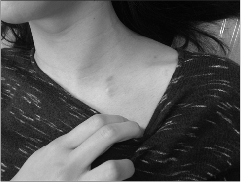
Figure 7 Photograph showing surgical wound after percutaneous Steinmann pin fixation.
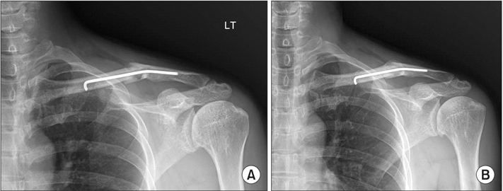
Figure 8 (A) Radiograph showing the bended Steinmann pin due to slip down. (B) Radiograph from postoperative 12 weeks shows bone union and remodeling without any other procedure.
Reference
-
1. Johnson EW Jr, Collins HR. Nonunion of the clavicle. Arch Surg. 1963; 87:963–966.2. Neer CS 2nd. Nonunion of the clavicle. J Am Med Assoc. 1960; 172:1006–1011.3. Paffen PJ, Jansen EW. Surgical treatment of clavicular fractures with Kirschner wires: a comparative study. Arch Chir Neerl. 1978; 30:43–53.4. Ljunggren AE. Clavicular function. Acta Orthop Scand. 1979; 50:261–268.5. Rowe CR. An atlas of anatomy and treatment of midclavicular fractures. Clin Orthop Relat Res. 1968; 58:29–42.6. Hill JM, McGuire MH, Crosby LA. Closed treatment of displaced middle-third fractures of the clavicle gives poor results. J Bone Joint Surg Br. 1997; 79:537–539.7. Baumgaertel F, Buhl M, Rahn BA. Fracture healing in biological plate osteosynthesis. Injury. 1998; 29:Suppl 3. C3–C6.8. Perren SM. Backgrounds of the technology of internal fixators. Injury. 2003; 34:Suppl 2. B1–B3.9. Perren SM. Evolution of the internal fixation of long bone fractures. The scientific basis of biological internal fixation: choosing a new balance between stability and biology. J Bone Joint Surg Br. 2002; 84:1093–1110.10. Robinson CM. Fractures of the clavicle in the adult. Epidemiology and classification. J Bone Joint Surg Br. 1998; 80:476–484.11. Beaton DE, Katz JN, Fossel AH, Wright JG, Tarasuk V, Bombardier C. Measuring the whole or the parts? Validity, reliability, and responsiveness of the disabilities of the arm, shoulder and hand outcome measure in different regions of the upper extremity. J Hand Ther. 2001; 14:128–146.12. Boehm D, Wollmerstedt N, Doesch M, Handwerker M, Mehling E, Gohlke F. Development of a questionnaire based on the Constant-Murley-Score for self-evaluation of shoulder function by patients. Unfallchirurg. 2004; 107:397–402.13. Kang KS, Ahn JI, Oh HY, Kang YS, Lee SJ. Clinical study of clavicle fractures. J Korean Orthop Assoc. 1984; 19:367–372.14. Eskola A, Vainionpää S, Myllynen P, Pätiälä H, Rokkanen P. Surgery for ununited clavicular fracture. Acta Orthop Scand. 1986; 57:366–367.15. Rockwood CA, Williams GR, Young DC. Acromioclavicular injuries. In : Rockwood CA, Green DP, Bucholz RW, Heckman JD, editors. Fractures in adults. Vol I. 4th ed. Philadelphia, PA: Lippincott-Raven;1996. p. 1109–1155.16. Robinson CM, Court-Brown CM, McQueen MM, Wakefield AE. Estimating the risk of nonunion following nonoperative treatment of a clavicular fracture. J Bone Joint Surg Am. 2004; 86:1359–1365.17. Frymoyer JW. Orthopaedic knowledge update. Am Acad Orthop Surg. 1993; 25:290.18. Böstman O, Manninen M, Pihlajamäki H. Complications of plate fixation in fresh displaced midclavicular fractures. J Trauma. 1997; 43:778–783.19. Post M. Current concepts in the treatment of fractures of the clavicle. Clin Orthop Relat Res. 1989; (245):89–101.20. Neviaser RJ, Neviaser JS, Neviaser TJ, Neviaser JS. A simple technique for internal fixation of the clavicle. A long term evaluation. Clin Orthop Relat Res. 1975; (109):103–107.21. Harnroongroj T, Jeerathanyasakun Y. Intramedullary pin fixation in clavicular fractures: a study comparing the use of small and large pins. J Orthop Surg (Hong Kong). 2000; 8:7–11.22. Wood VE. The results of total claviculectomy. Clin Orthop Relat Res. 1986; (207):186–190.23. Poigenfürst J, Reiler T, Fischer W. Plating of fresh clavicular fractures. Experience with 60 operations. Unfallchirurgie. 1988; 14:26–37.24. Stanley D, Norris SH. Recovery following fractures of the clavicle treated conservatively. Injury. 1988; 19:162–164.
- Full Text Links
-
- Actions
-
Cited
- CITED
-
- Close
- Share
-
- Similar articles
-
- Surgical Techniques for Percutaneous Reduction by Towel Clips and Percutaneous Intramedullary Fixation with Steinmann Pins for Clavicle Shaft Fractures
- Treatment of the Fracture of the Middle Third of Clavicle by Intramedullary Threaded Steinmann Pin Fixation
- Comparison of Results in Two Operative Treatments for Clavicle Shaft Fractures in Adult: Comparison of Results between Open Reduction and Internal Fixation with the Plate and Percutaneous Reduction by Towel Clip and Intramedullary Fixation with Steinmann Pin
- Intramedullary Fixation of Clavicle Fracture Percutaneously Reduced By Towel Clip
- Intramedullary Fixation in the Fracture of the Shaft of the Clavicle by Threaded Kirschner Wire
