Complication and management of implant-assisted removable partial denture with distal extension: a clinical report
- Affiliations
-
- 1Department of Prosthodontics, Institute of Oral Bio-Science, School of Dentistry, Chonbuk National University and Research Institute of Clinical Medicine of Chonbuk National University-Biomedical Research Institute of Chonbuk National University Hospital,
- 2Department of Dentistry, School of Medicine, Eulji University, Daejeon, Republic of Korea.
- KMID: 2369075
- DOI: http://doi.org/10.14368/jdras.2016.32.4.338
Abstract
- Implant supported removable partial denture (ISRPD) using the implants enables favorable rehabilitation by complementing biomechanical limitations of the conventional removable partial denture (RPD). However, continuous recall check is necessary for evaluation of the mechanical and biological complications to ensure good long-term prognosis of ISRPD. This clinical report describes the complication and management in patient of Kennedy class I edentulism with ISRPD using healing abutment. The wear and fracture of healing abutment occurred at 36 months after delivery. So, healing abutment was replaced by connecting Locator® abutment for altering into the implant retained partial overdenture.
MeSH Terms
Figure
-
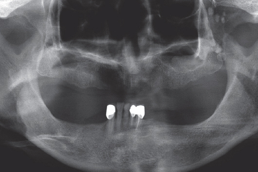
Fig. 1 Initial panoramic radiograph.
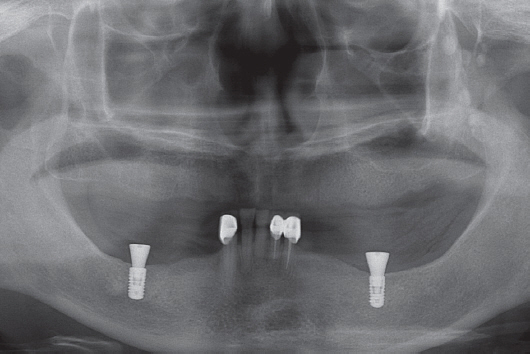
Fig. 2 Postoperative panoramic radiograph. Two short implants were placed in mandibular posterior area.
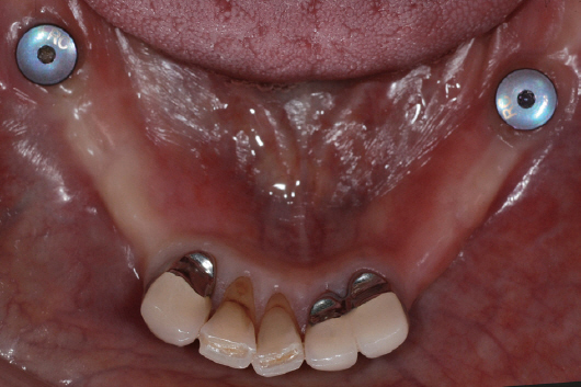
Fig. 3 Anterior surveyed prostheses were cemented. No inflammatory signs were observed around healing abutment.
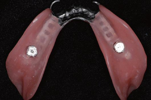
Fig. 4 Definitive removable partial denture was fabricated. Metal framework was adjusted to minimum contact with the healing abutment.
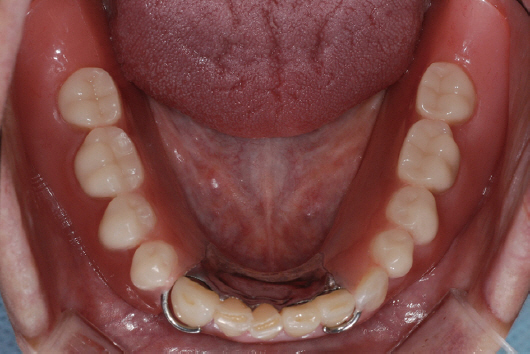
Fig. 5 Delivery of removable partial denture.
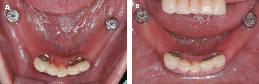
Fig. 6 (A) Occlusal view at 18 months after delivery. Mechanical wear was observed on flat surface of healing abutment, (B) At 30 months after delivery.
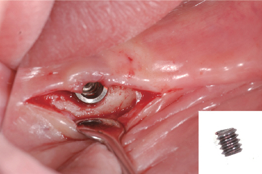
Fig. 7 Fragment of healing abutment was removed on mandibular right implant. Fracture of implant fixture was not observed.
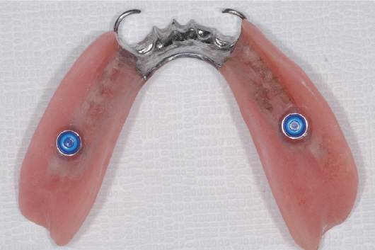
Fig. 8 Old prosthesis was repaired. Blue nylon patrix was inserted.
Reference
-
References
1. Kratochvil FJ, Caputo AA. Photoelastic analysis of pressure on teeth and bone supporting removable partial dentures. J Prosthet Dent. 1974; 32:52–61. DOI: 10.1016/0022-3913(74)90098-5.2. Shahmiri R, Aarts JM, Bennani V, Das R, Swain MV. Strain distribution in a Kennedy Class I implant assisted removable partial denture under various loading conditions. Int J Dent. 2013; 2013:351279. DOI: 10.1155/2013/351279. PMID: 23737788. PMCID: PMC3657413.3. Kuzmanovic DV, Payne AG, Purton DG. Distal implants to modify the Kennedy classification of a removable partial denture: a clinical report. J Prosthet Dent. 2004; 92:8–11. DOI: 10.1016/j.prosdent.2004.04.010. PMID: 15232557.4. Wismeijer D, Tawse-Smith A, Payne AG. Multicentre prospective evaluation of implant-assisted mandibular bilateral distal extension removable partial dentures: patient satisfaction. Clin Oral Implants Res. 2013; 24:20–7. DOI: 10.1111/j.1600-0501.2011.02367.x. PMID: 22111809.5. Chikunov I, Doan P, Vahidi F. Implant-retained partial overdenture with resilient attachments. J Prosthodont. 2008; 17:141–8. DOI: 10.1111/j.1532-849X.2007.00261.x. PMID: 18005337.6. Ohkubo C, Kobayashi M, Suzuki Y, Hosoi T. Effect of implant support on distal-extension removable partial dentures: in vivo assessment. Int J Oral Maxillofac Implants. 2008; 23:1095–101. PMID: 19216279.7. de Freitas RF, de Carvalho Dias K, da Fonte Porto Carreiro A, Barbosa GA, Ferreira MA. Mandibular implant-supported removable partial denture with distal extension: a systematic review. J Oral Rehabil. 2012; 39:791–8. DOI: 10.1111/j.1365-2842.2012.02326.x. PMID: 22882547.8. McGarry TJ, Nimmo A, Skiba JF, Ahlstrom RH, Smith CR, Koumjian JH, Arbree NS. Classification system for partial edentulism. J Prosthodont. 2002; 11:181–93. DOI: 10.1053/jopr.2002.126094. DOI: 10.1053/jpro.2002.126094. PMID: 12237799.9. Cunha LD, Pellizzer EP, Verri FR, Pereira JA. Evaluation of the influence of location of osseointegrated implants associated with mandibular removable partial dentures. Implant Dent. 2008; 17:278–87. DOI: 10.1097/ID.0b013e31818363b2. PMID: 18784528.10. Verri FR, Pellizzer EP, Rocha EP, Pereira JA. Influence of length and diameter of implants associated with distal extension removable partial dentures. Implant Dent. 2007; 16:270–80. J Dent Rehabil Appl Sci. 2016; 32(4):338–44. 343.11. Mitrani R, Brudvik JS, Phillips KM. Posterior implants for distal extension removable prostheses: a retrospective study. Int J Periodontics Restorative Dent. 2003; 23:353–9. PMID: 12956479.12. Pellizzer EP, Verri FR, Falcón-Antenucci RM, Goiato MC, Gennari Filho H. Evaluation of different retention systems on a distal extension removable partial denture associated with an osseointegrated implant. J Craniofac Surg. 2010; 21:727–34. DOI: 10.1097/SCS.0b013e3181d8098a. PMID: 20485037.13. Grossmann Y, Nissan J, Levin L. Clinical effectiveness of implant-supported removable partial dentures: a review of the literature and retrospective case evaluation. J Oral Maxillofac Surg. 2009; 67:1941–6. DOI: 10.1016/j.joms.2009.04.081. PMID: 19686933.14. ELsyad MA, Omran AO, Fouad MM. Strains around abutment teeth with different attachments used for implant-assisted distal extension partial overdentures: an in vitro study. J Prosthodont. 2015; Sep. 29. doi:10.1111/jopr.12370. [Epub ahead of print]. DOI: 10.1111/jopr.12370.15. el Charkawi HG, Zekry KA, el Wakad MT. Stress analysis of different osseointegrated implants supporting a distal extension prosthesis. J Prosthet Dent. 1994; 72:614–22. DOI: 10.1016/0022-3913(94)90294-1.16. Mahrous AI, Aldawash HA, Soliman TA, Banasr FH, Abdelwahed A. Implant supported distal extension over denture retained by two types of attachments. A comparative radiographic study by cone beam computed tomography. J Int Oral Health. 2015; 7:5–10.
- Full Text Links
-
- Actions
-
Cited
- CITED
-
- Close
- Share
-
- Similar articles
-
- Restoration of bilateral distal extension removable partial denture using a fixed implant prosthesis in unilateral partial edentulous patient: A case report
- Implant-assisted removable partial denture restoration in a partially edentulous patient with a single remaining tooth: a case report
- Implant assisted removable partial denture with implant surveyed crown: A 20-month follow-up case report
- Rehabilitation of maxillary partial edentulous patients using implant assisted removable partial denture
- Implant-assisted removable partial denture in a maxillary edentulous patient: A case report
