Laterally positioned flap using subepithelial connective tissue graft for iatrogenic gingival recession treatment
- Affiliations
-
- 1Department of Periodontology, College of Dentistry, Dankook University, Cheonan, Republic of Korea. periolee85@gmail.com
- KMID: 2369074
- DOI: http://doi.org/10.14368/jdras.2016.32.4.330
Abstract
- Gingival recession could occur after orthodontic or endodontic treatment. This could influence not only functional and aesthetic problem, but also patient's treatment satisfaction. There are a lot of techniques for preventing gingival recession, but laterally positioned flap with subepithelial connective tissue graft could get definite advantages such as increase of keratinized gingival width and harmonious color match between graft tissue and surrounding tissue. Clinician should select a right patient case and diagnosis with clinical and radiography exam. In a surgical procedure, getting enough amounts of subepithelial connective tissue and flap coverage should be taken into consideration. The clinical outcomes in this case report shows laterally positioned flap with subepithelial connective tissue graft could be a treatment with predictive outcome.
MeSH Terms
Figure
-
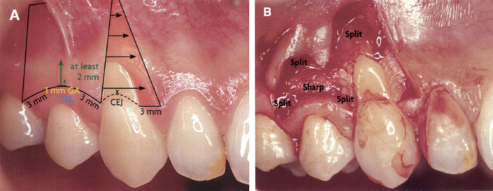
Fig. 1 (A) Donor & recipient site design, (B) Mix-thickness flap. CEJ, cementoenamel junction; Split, split-thickness flap; Sharp, sharp horizontal incision & full-thickness flap.
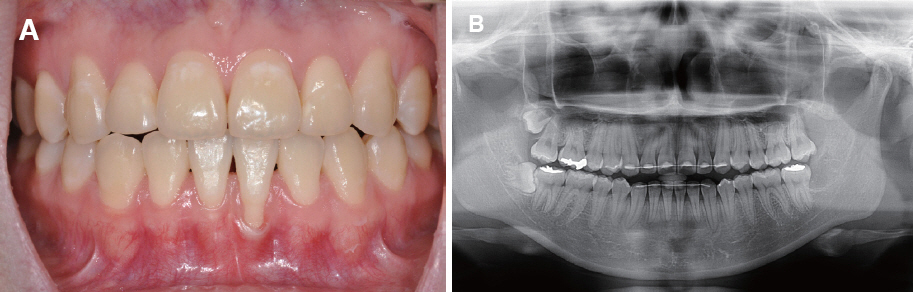
Fig. 2 (A) Initial aspect of Case I showing Miller Class II localized gingival recession at the mandibular left central incisor, (B) Panoramic radiography of Case I.
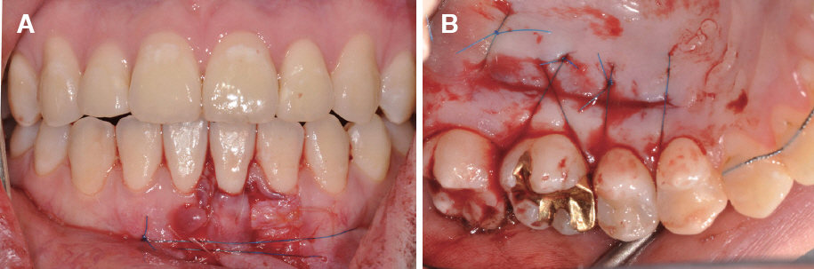
Fig. 3 Clinical view of surgical procedure. (A) A lateral flap was positioned over the connective tissue graft and sutured, (B) Palatal donor site was sutured.
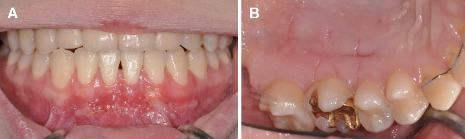
Fig. 4 Postoperative clinical view after 15 days. (A) Stable healing observed at recipient site and (B) palatal donor site.
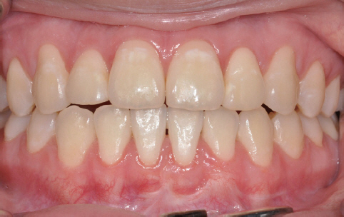
Fig. 5 After 3 months clinical view of Case I.
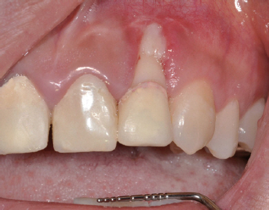
Fig. 6 Initial aspect of Case II showing Miller Class II localized gingival recession with sequestrum at the maxillary left lateral incisor.
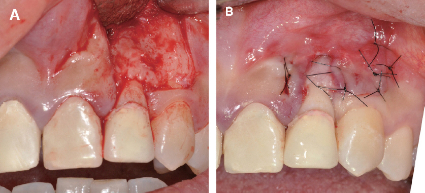
Fig. 7 Clinical view of surgical procedure for sequestrectomy. (A) Sequestrum observed below the recessed gingival margin, (B) Suture after trimming sequestrum.
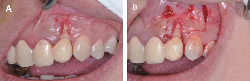
Fig. 8 Clinical view of surgical procedure. (A) Showing 3 mm gingival receesion, (B) Partial thickness flap incision making for recitient site.
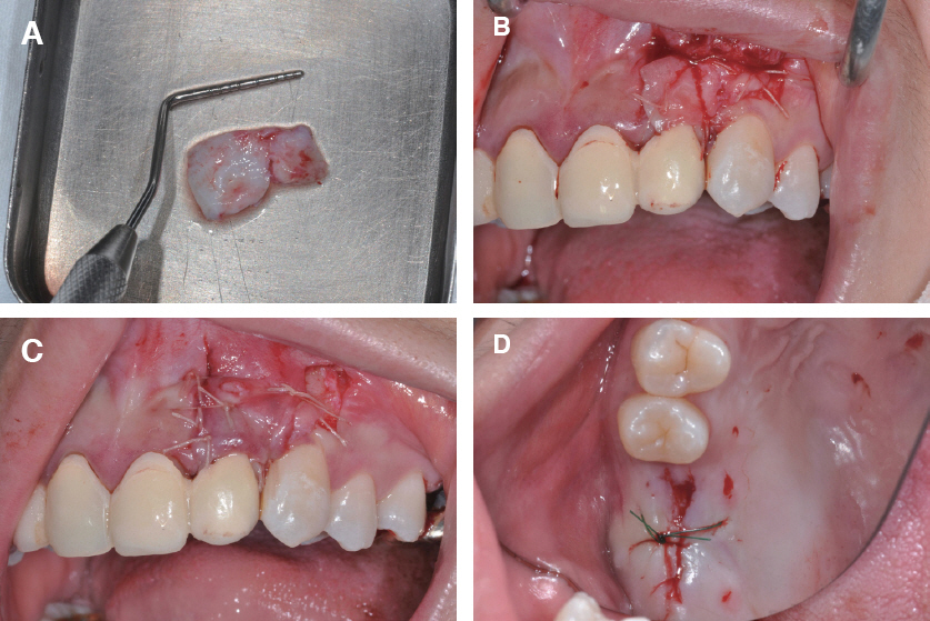
Fig. 9 Clinical view of surgical procedure. (A) Subepithelial connective tissue grafting from edentulous ridge, (B) The graft tissue positioned at recipient site, (C) A lateral flap was positioned over the connective tissue graft and sutured, (D) Edentulous donor site was sutured.
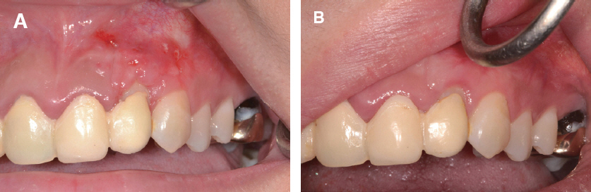
Fig. 10 (A) Postoperative clinical view after 15 days and (B) after 3 months. 1 mm gingival recession remained.
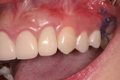
Fig. 11 After 1 year clinical view of Case II.
Reference
-
References
1. Hall WB. Present status of soft tissue grafting. J Periodontol. 1977; 48:587–97. DOI: 10.1902/jop.1977.48.9.587. PMID: 333092.2. Melsen B, Allais D. Factors of importance for the development of dehiscences during labial movement of mandibular incisors: a retrospective study of adult orthodontic patients. Am J Orthod Dentofacial Orthop. 2005; 127:552–61. DOI: 10.1016/j.ajodo.2003.12.026. PMID: 15877035.3. Seltzer S, Sinai I, August D. Periodontal effects of root perforations before and during endodontic procedures. J Dent Res. 1970; 49:332–9. DOI: 10.1177/00220345700490022301. PMID: 4984781.4. Miller PD Jr. A classification of marginal tissue recession. Int J Periodontics Restorative Dent. 1985; 5:8–13. PMID: 3858267.5. Grupe HE, Warren RF Jr. Repair of gingival defects by a sliding flap operation. J Periodontol. 1956; 27:92–5. DOI: 10.1902/jop.1956.27.2.92.6. Grupe HE. Modified technique for the sliding flap operation. J Periodontol. 1966; 37:491–5. DOI: 10.1902/jop.1966.37.6.491. PMID: 5224017.7. Lee CT, Chang PC, Touchan N, Royzman D. Root coverage with a modified laterally positioned flap combined with a subepithelial connective tissue graft in advanced recession. J Periodontal Implant Sci. 2014; 44:300–6. DOI: 10.5051/jpis.2014.44.6.300. PMID: 25568811. PMCID: PMC4284379.8. Zucchelli G. Mucogingival esthetic surgery. 1st ed. Milano;Quintessenza Edizioni;. 2013; 330–78.9. Jahnke PV, Sandifer JB, Gher ME, Gray JL, Richardson AC. Thick free gingival and connective tissue autografts for root coverage. J Periodontol. 1993; 64:315–22. DOI: 10.1902/jop.1993.64.4.315. PMID: 8483096.10. Carnio J, Neiva RF. Mineral trioxide aggregate and subepithelial connective tissue graft for treatment of iatrogenic gingival recession: long-term results. Int J Periodontics Restorative Dent. 2014; 34:71–7. DOI: 10.11607/prd.1674. PMID: 24396836.11. Jung UW, Kim CS, Choi SH, Kim S. Gingival coverage of iatrogenically denuded labial bone resulting from thermal trauma. Int J Periodontics Restorative Dent. 2013; 33:635–9. DOI: 10.11607/prd.1024. PMID: 23998159.12. Ruben MP. Rationale for the employment of laterally positioned flaps and free autogenous gingival grafts in periodontics (I). Quintessence Int Dent Dig. 1978; 9:57–61.13. Ruben MP. Rationale for the employment of laterally positioned flaps and free autogenous gingival grafts in periodontics (II). Quintessence Int Dent Dig. 1978; 9:53–7. PMID: 288094.14. Hall WB. Gingival augmentation/mucogingival surgery. Nevins M, Becker W, Kornman K, editors. Proceedings of the World Workshop in Clinical Periodontics. Chicago: American Academy of Periodontology;1989. VII:p. 1–21. PMCID: PMC324834.15. Santana RB, Furtado MB, Mattos CM, de Mello Fonseca E, Dibart S. Clinical evaluation of singlestage advanced versus rotated flaps in the treatment of gingival recessions. J Periodontol. 2010; 81:48592. DOI: 10.1902/jop.2010.090237. PMID: 20367091.16. Zucchelli G, Cesari C, Amore C, Montebugnoli L, De Sanctis M. Laterally moved, coronally advanced flap: a modified surgical approach for isolated recession-type defects. J Periodontol. 2004; 75:173441. DOI: 10.1902/jop.2004.75.12.1734. PMID: 15732880.17. Zuhr O, Bäumer D, Hürzeler M. The addition of soft tissue replacement grafts in plastic periodontal and implant surgery: critical elements in design and execution. J Clin Periodontol. 2014; 41(suppl 15):S123–42. DOI: 10.1111/jcpe.12185. PMID: 24640997.18. Yotnuengnit P, Promsudthi A, Teparat T, Laohapand P, Yuwaprecha W. Relative connective tissue graft size affects root coverage treatment outcome in the envelope procedure. J Periodontol. 2004; 75:886–92. DOI: 10.1902/jop.2004.75.6.886. PMID: 15295957.19. Langer B, Langer L. Subepithelial connective tissue graft technique for root coverage. J Periodontol. 1985; 56:715–20. DOI: 10.1902/jop.1985.56.12.715. PMID: 3866056.20. Miller PD Jr. Root coverage with the free gingival graft. Factors associated with incomplete coverage. J Periodontol. 1987; 58:674–81. DOI: 10.1902/jop.1987.58.10.674. PMID: 3478464.
- Full Text Links
-
- Actions
-
Cited
- CITED
-
- Close
- Share
-
- Similar articles
-
- Root coverage using laterally positioned flap and subepithelial connective tissue graft for the treatment of the isolated recession defects on mandibular anterior teeth: case report
- Root coverage with a modified laterally positioned flap combined with a subepithelial connective tissue graft in advanced recession
- Root coverage with subeptithelial connective tissue grafts
- The evaluation of clinical outcomes on various procedures using subepithelial connective tissue graft for coverage of gingival recession
- Comparative Evaluation of Connective Tissue Graft with Pouch/ Tunnel Technique versus Connective Tissue Graft with Coronally Advanced Tunnel Flap for the Treatment of Maxillary Recession Cases in Severe Periodontitis
