J Periodontal Implant Sci.
2017 Feb;47(1):30-40. 10.5051/jpis.2017.47.1.30.
Comparisons of the diagnostic accuracies of optical coherence tomography, micro-computed tomography, and histology in periodontal disease: an ex vivo study
- Affiliations
-
- 1Department of Periodontology, Research Institute for Periodontal Regeneration, Yonsei University College of Dentistry, Seoul, Korea. drjew@yuhs.ac
- 2Intelligence R&D Laboratory, LG Electronics, Seoul, Korea.
- 3Division of Anatomy and Developmental Biology, Department of Oral Biology, Human Identification Research Center, Yonsei University College of Dentistry, Seoul, Korea.
- KMID: 2368885
- DOI: http://doi.org/10.5051/jpis.2017.47.1.30
Abstract
- PURPOSE
Optical coherence tomography (OCT) is a noninvasive diagnostic technique that may be useful for both qualitative and quantitative analyses of the periodontium. Micro-computed tomography (micro-CT) is another noninvasive imaging technique capable of providing submicron spatial resolution. The purpose of this study was to present periodontal images obtained using ex vivo dental OCT and to compare OCT images with micro-CT images and histologic sections.
METHODS
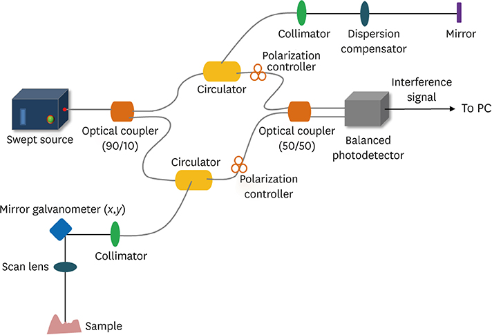
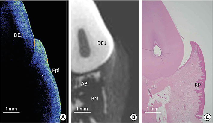
Images of ex vivo canine periodontal structures were obtained using OCT. Biologic depth measurements made using OCT were compared to measurements made on histologic sections prepared from the same sites. Visual comparisons were made among OCT, micro-CT, and histologic sections to evaluate whether anatomical details were accurately revealed by OCT.
RESULTS
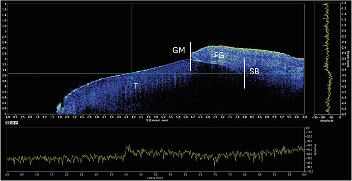
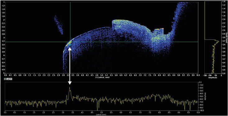
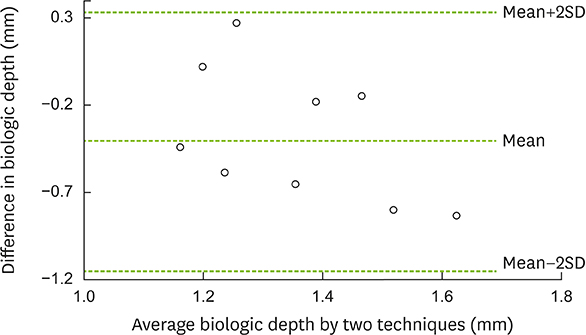
The periodontal tissue contour, gingival sulcus, and the presence of supragingival and subgingival calculus could be visualized using OCT. OCT was able to depict the surface topography of the dentogingival complex with higher resolution than micro-CT, but the imaging depth was typically limited to 1.2-1.5 mm. Biologic depth measurements made using OCT were a mean of 0.51 mm shallower than the histologic measurements.
CONCLUSIONS
Dental OCT as used in this study was able to generate high-resolution, cross-sectional images of the superficial portions of periodontal structures. Improvements in imaging depth and the development of an intraoral sensor are likely to make OCT a useful technique for periodontal applications.
Figure
Reference
-
1. Brown LJ, Löe H. Prevalence, extent, severity and progression of periodontal disease. Periodontol 2000. 1993; 2:57–71.
Article2. Petersen PE. The World Oral Health Report 2003: continuous improvement of oral health in the 21st century--the approach of the WHO Global Oral Health Programme. Community Dent Oral Epidemiol. 2003; 31:Suppl 1. 3–23.3. Burt B. Research, Science and Therapy Committee of the American Academy of Periodontology. Position paper: epidemiology of periodontal diseases. J Periodontol. 2005; 76:1406–1419.4. Petersen PE, Ogawa H. The global burden of periodontal disease: towards integration with chronic disease prevention and control. Periodontol 2000. 2012; 60:15–39.
Article5. Armitage GC. Research, Science and Therapy Committee of the American Academy of Periodontology. Diagnosis of periodontal diseases. J Periodontol. 2003; 74:1237–1247.6. Pihlstrom BL. Measurement of attachment level in clinical trials: probing methods. J Periodontol. 1992; 63:1072–1077.
Article7. Mayfield L, Bratthall G, Attström R. Periodontal probe precision using 4 different periodontal probes. J Clin Periodontol. 1996; 23:76–82.
Article8. Molander B, Ahlqwist M, Gröndahl HG, Hollender L. Comparison of panoramic and intraoral radiography for the diagnosis of caries and periapical pathology. Dentomaxillofac Radiol. 1993; 22:28–32.
Article9. Jeffcoat MK, Wang IC, Reddy MS. Radiographic diagnosis in periodontics. Periodontol 2000. 1995; 7:54–68.
Article10. Croucher PI, Garrahan NJ, Compston JE. Assessment of cancellous bone structure: comparison of strut analysis, trabecular bone pattern factor, and marrow space star volume. J Bone Miner Res. 1996; 11:955–961.
Article11. Otis LL, Colston BW Jr, Everett MJ, Nathel H. Dental optical coherence tomography: a comparison of two in vitro systems. Dentomaxillofac Radiol. 2000; 29:85–89.
Article12. Mota CC, Fernandes LO, Cimões R, Gomes AS. Non-invasive periodontal probing through fourier-domain optical coherence tomography. J Periodontol. 2015; 86:1087–1094.
Article13. Fried D, Glena RE, Featherstone JD, Seka W. Nature of light scattering in dental enamel and dentin at visible and near-infrared wavelengths. Appl Opt. 1995; 34:1278–1285.
Article14. Feldchtein F, Gelikonov V, Iksanov R, Gelikonov G, Kuranov R, Sergeev A, et al. In vivo OCT imaging of hard and soft tissue of the oral cavity. Opt Express. 1998; 3:239–250.15. Brezinski ME, Tearney GJ, Bouma BE, Izatt JA, Hee MR, Swanson EA, et al. Optical coherence tomography for optical biopsy. Properties and demonstration of vascular pathology. Circulation. 1996; 93:1206–1213.
- Full Text Links
- Actions
-
Cited
- CITED
-
- Close
- Share
- Similar articles
-
- Micro-computed tomography analysis of changes in the periodontal ligament and alveolar bone proper induced by occlusal hypofunction of rat molars
- Availability of Optical Coherence Tomography in Diagnosis and Classification of Choroidal Neovascularization
- A Newly Developed Stent Thrombus Related to Optical Coherence Tomography
- Optical coherence tomography angiography in preclinical neuroimaging
- The Use fulness of OCT[Optical Coherence Tomography]for the Diagnosis of Central Serous Choriore tinopathy

