Ann Dermatol.
2014 Jun;26(3):419-421.
Small Cell Melanoma
- Affiliations
-
- 1Department of Dermatology, Ajou University School of Medicine, Suwon, Korea. maychan@ajou.ac.kr
Abstract
- No abstract available.
MeSH Terms
Figure
-

Fig. 1 A solitary black patch on sutured skin and irregular brownish to bluish small patches beside the excised area were observed on the patient's right sole.
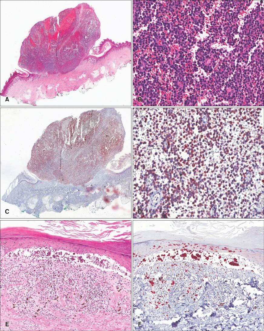
Fig. 2 The excised nodular lesion was composed of small cells. The round small cells were immunoreactive to Melan-A (A~D). Beside the nodular lesion, a black patch with lentiginous proliferation of melanocytes and showing reactivity to Melan-A was observed (E, F). (A) H&E, ×4; (B) H&E, ×200; (C) immunohistochemical stain, ×4; (D) immunohistochemical stain, ×200; (E) H&E, ×100; (F) immunohistochemical stain, ×100.
Reference
-
1. Eyden B, Moss J, Shore I, Banerjee SS. Metastatic small cell malignant melanoma: a case requiring immunoelectron-microscopy for the demonstration of lattice-deficient melanosomes. Ultrastruct Pathol. 2005; 29:71–78.
Article2. Nakhleh RE, Wick MR, Rocamora A, Swanson PE, Dehner LP. Morphologic diversity in malignant melanomas. Am J Clin Pathol. 1990; 93:731–740.
Article3. Magro CM, Crowson AN, Mihm MC. Unusual variants of malignant melanoma. Mod Pathol. 2006; 19:Suppl 2. S41–S70.
Article4. Lever WF, Elder DE. Lever's histopathology of the skin. 10th ed. Philadelphia: Wolters Kluwer Health/Lippincott Williams & Wilkins;2009. p. xix. p. 1257.
- Full Text Links
-
- Actions
-
Cited
- CITED
-
- Close
- Share
-
- Similar articles
-
- A Case of Melanoma in situ Arising in a Small Congenital Melanocytic Nevus
- Growth and Characterization of the Uveal Melanoma in Vitro
- Malignant Melanoma Developed from Giant Congenital pigmented Nevus: Report of a Case
- A Case of Malignant Melanoma Arising in Small Congenital Melanocytic Nevus
- Malignant Melanoma on Congenital Melanocytic Nevus
