Ann Dermatol.
2014 Jun;26(3):403-404.
Clear Cell Hidradenoma on the Palm
- Affiliations
-
- 1Department of Dermatology, Samsung Medical Center, Sungkyunkwan University School of Medicine, Seoul, Korea. dylee@skku.edu
- 2Department of Dermatology, Kangbuk Samsung Hospital, Sungkyunkwan University School of Medicine, Seoul, Korea.
Abstract
- No abstract available.
MeSH Terms
Figure
-
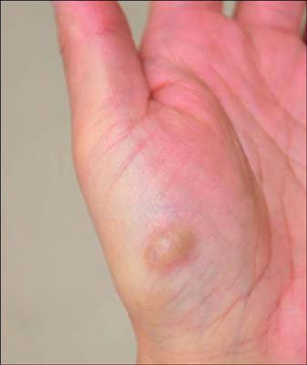
Fig. 1 A brownish nodule on the thenar area of the palm.
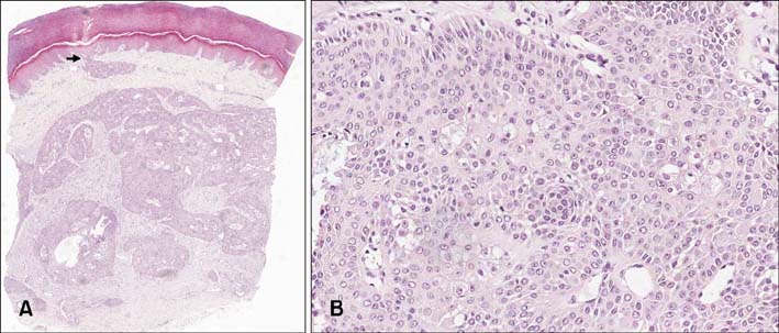
Fig. 2 (A) A circumscribed dermal neoplasm with mostly solid areas and some cystic areas with myxoid stroma. An epidermal connection was observed (black arrow; H&E, ×40, scanning view). (B) Clusters of clear cells were observed (H&E, ×100).
Reference
-
1. Hernandez-Perez E, Cestoni-Parducci R. Nodular hidradenoma and hidradenocarcinoma. A 10-year review. J Am Acad Dermatol. 1985; 12:15–20.2. Gianotti R, Alessi E. Clear cell hidradenoma associated with the folliculo-sebaceous-apocrine unit. Histologic study of five cases. Am J Dermatopathol. 1997; 19:351–357.
Article3. Nandeesh BN, Rajalakshmi T. A study of histopathologic spectrum of nodular hidradenoma. Am J Dermatopathol. 2012; 34:461–470.
Article4. Battistella M, Langbein L, Peltre B, Cribier B. From hidroacanthoma simplex to poroid hidradenoma: clinicopathologic and immunohistochemic study of poroid neoplasms and reappraisal of their histogenesis. Am J Dermatopathol. 2010; 32:459–468.
Article5. Langbein L, Cribier B, Schirmacher P, Praetzel-Wunder S, Peltre B, Schweizer J. New concepts on the histogenesis of eccrine neoplasia from keratin expression in the normal eccrine gland, syringoma and poroma. Br J Dermatol. 2008; 159:633–645.
Article
