Ann Dermatol.
2014 Jun;26(3):399-400.
Acute Generalized Exanthematous Pustulosis Induced by Parvovirus B19 Infection
- Affiliations
-
- 1Department of Dermatology, Busan Paik Hospital, Inje University College of Medicine, Busan, Korea. derma09@hanmail.net
- 2Sinsa Theme Dermatology Clinic, Seoul, Korea.
Abstract
- No abstract available.
Figure
-
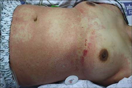
Fig. 1 Multiple nonfollicular pustules with surround erythema on the trunk.
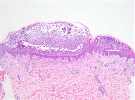
Fig. 2 Histopathologic findings shows subcorneal pustules in the epidermis, perivascular and interstitial inflammatory cells infiltration in the dermis. The inflammatory cells are comprised of lymphohistiocytes, neutrophils and a few eosinophils (H&E, ×40).
Reference
-
1. Sidoroff A, Dunant A, Viboud C, Halevy S, Bavinck JN, Naldi L, et al. Risk factors for acute generalized exanthematous pustulosis (AGEP)-results of a multinational case-control study (EuroSCAR). Br J Dermatol. 2007; 157:989–996.
Article2. Vafaie J, Schwartz RA. Parvovirus B19 infections. Int J Dermatol. 2004; 43:747–749.
Article3. Perceau G, Derancourt C, Salmon-Ehr V, Durlach A, Raclot P, Bredard V, et al. Acute generalized exanthematous pustulosis in hypercalcemia. Ann Dermatol Venereol. 2000; 127:1090–1093.4. Ofuji S, Yamamoto O. Acute generalized exanthematous pustulosis associated with a human parvovirus B19 infection. J Dermatol. 2007; 34:121–123.
Article5. Fernando SL. Acute generalised exanthematous pustulosis. Australas J Dermatol. 2012; 53:87–92.
Article
- Full Text Links
-
- Actions
-
Cited
- CITED
-
- Close
- Share
-
- Similar articles
-
- Acute Generalized Exanthematous Pustulosis with Hemodynamic Instability Induced by Ingestion of Lacquer Chicken
- A Case of Acute Generalized Exanthematous Pustulosis Possibly Induced by Ritodrine
- A Serial Occurrence of Acute Generalized Exanthematous Pustulosis in Childhood Siblings
- Acute Generalized Exanthematous Pustulosis
- Acute generalized exanthematous pustulosis induced by terbinafine
