Sebaceous Carcinoma of the Suprapubic Area in a Liver Transplant Recipient
- Affiliations
-
- 1Department of Plastic and Reconstructive Surgery, College of Medicine, The Catholic University of Korea, Seoul, Korea. rhie@catholic.ac.kr
Abstract
- Sebaceous carcinoma is a very rare and potentially aggressive carcinoma originating from the epithelial lining of the sebaceous gland. More than 70% of all cases are in the head and neck region, especially the periorbita; therefore, they are classified into ocular and extraocular sebaceous carcinoma. The reported risk factors are advanced age, male sex, previous irradiation, and genetic predisposition for Muir-Torre syndrome. The current case is of sebaceous carcinoma found in the suprapubic area of a 67-year-old male patient who had received liver transplantation 6 years before, and had been receiving oral tacrolimus. Examination of the gastrointestinal system did not reveal any other malignancies. Although nonmelanoma skin cancers may occur as a complication after liver transplantation, there have been no previous reports of sebaceous carcinoma after liver transplantation. Furthermore, the sebaceous carcinoma in this case occurred in an uncommon location. We report this case along with a review of the literature.
MeSH Terms
Figure
-

Fig. 1 A pink, well-circumscribed suprapubic mass with a diameter of 3 cm. Ulceration is seen on its right upper surface.

Fig. 2 Computed tomography showing a well-defined mass in the skin of the lower abdominal wall.
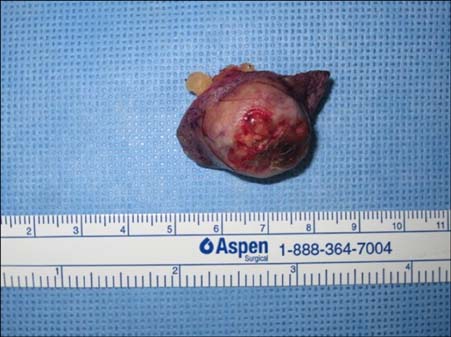
Fig. 3 The specimen after excision with 5-mm margins.
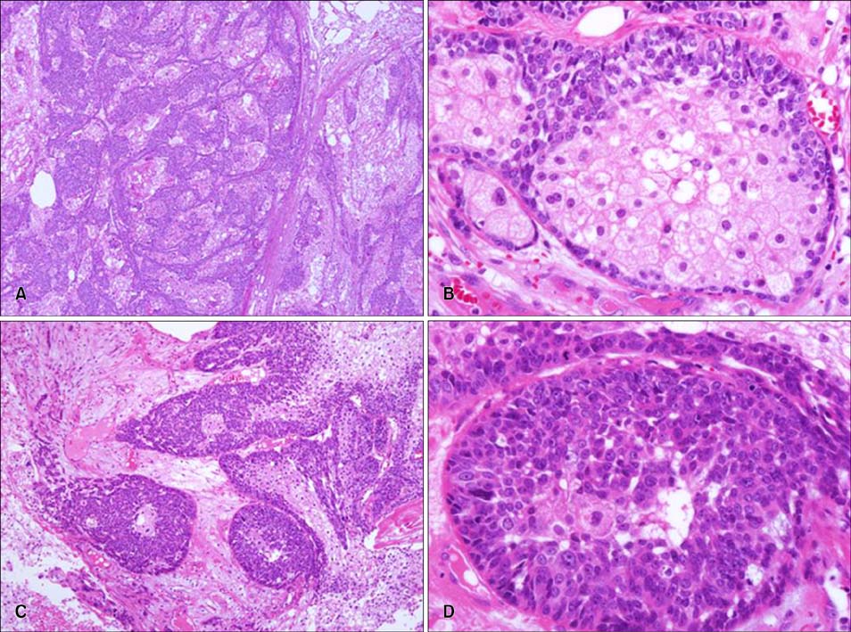
Fig. 4 Histopathological findings. (A) The well-differentiated areas show small lobular aggregations of sebocytes (H&E, ×40). (B) The margins consist of basaloid cells and the central portions are fully differentiated vacuolated sebocytes, as in normal sebaceous glands (H&E, ×400). (C) Poorly differentiated areas have high cellularity and invasion into the surrounding tissue (H&E, ×40). (D) Atypism (nuclear pleomorphism, prominent nucleoli, high nucleus-to-cell ratio) is seen, and mitosis is observed, with almost no differentiation into normal sebocytes (H&E, ×400).
Reference
-
1. Jo MS, Kwon KH, Shin HK, Choe J, Jang TJ. Sebaceous carcinoma arising from the nevus sebaceous. Arch Plast Surg. 2012; 39:431–433.
Article2. Dasgupta T, Wilson LD, Yu JB. A retrospective review of 1349 cases of sebaceous carcinoma. Cancer. 2009; 115:158–165.
Article3. Chao AN, Shields CL, Krema H, Shields JA. Outcome of patients with periocular sebaceous gland carcinoma with and without conjunctival intraepithelial invasion. Ophthalmology. 2001; 108:1877–1883.
Article4. Sakol PJ, Simons KB, McFadden PW, Harris GJ, Massaro BM, Koethe S. DNA flow cytometry of sebaceous cell carcinomas of the ocular adnexa: introduction to the technique in the evaluation of periocular tumors. Ophthal Plast Reconstr Surg. 1992; 8:77–87.5. Kivelä T, Asko-Seljavaara S, Pihkala U, Hovi L, Heikkonen J. Sebaceous carcinoma of the eyelid associated with retinoblastoma. Ophthalmology. 2001; 108:1124–1128.
Article6. Harwood CA, McGregor JM, Swale VJ, Proby CM, Leigh IM, Newton R, et al. High frequency and diversity of cutaneous appendageal tumors in organ transplant recipients. J Am Acad Dermatol. 2003; 48:401–408.
Article7. Kaminska EC, Iyengar V, Tsoukas M, Shea CR. Borderline sebaceous neoplasm in a renal transplant patient without Muir-Torre syndrome. J Cutan Pathol. 2013; 40:336–340.
Article8. Euvrard S, Morelon E, Rostaing L, Goffin E, Brocard A, Tromme I, et al. TUMORAPA Study Group. Sirolimus and secondary skin-cancer prevention in kidney transplantation. N Engl J Med. 2012; 367:329–339.
Article9. Gaskin BJ, Fernando BS, Sullivan CA, Whitehead K, Sullivan TJ. The significance of DNA mismatch repair genes in the diagnosis and management of periocular sebaceous cell carcinoma and Muir-Torre syndrome. Br J Ophthalmol. 2011; 95:1686–1690.
Article10. Nelson BR, Hamlet KR, Gillard M, Railan D, Johnson TM. Sebaceous carcinoma. J Am Acad Dermatol. 1995; 33:1–15. quiz 16-18.
Article11. Harwood CA, Swale VJ, Bataille VA, Quinn AG, Ghali L, Patel SV, et al. An association between sebaceous carcinoma and microsatellite instability in immunosuppressed organ transplant recipients. J Invest Dermatol. 2001; 116:246–253.
Article12. Guba M, Graeb C, Jauch KW, Geissler EK. Pro- and anti-cancer effects of immunosuppressive agents used in organ transplantation. Transplantation. 2004; 77:1777–1782.
Article13. Maluccio M, Sharma V, Lagman M, Vyas S, Yang H, Li B, et al. Tacrolimus enhances transforming growth factor-beta1 expression and promotes tumor progression. Transplantation. 2003; 76:597–602.
Article14. Euvrard S, Kanitakis J, Claudy A. Skin cancers after organ transplantation. N Engl J Med. 2003; 348:1681–1691.
Article15. Park HW, Hwang S, Ahn CS, Kim KH, Moon DB, Ha TY, et al. De novo malignancies after liver transplantation: incidence comparison with the Korean cancer registry. Transplant Proc. 2012; 44:802–805.
Article16. Buitrago W, Joseph AK. Sebaceous carcinoma: the great masquerader: emgerging concepts in diagnosis and treatment. Dermatol Ther. 2008; 21:459–466.17. Hata M, Koike I, Omura M, Maegawa J, Ogino I, Inoue T. Noninvasive and curative radiation therapy for sebaceous carcinoma of the eyelid. Int J Radiat Oncol Biol Phys. 2012; 82:605–611.
Article18. Sawyer AR, McGoldrick RB, Mackey S, Powell B, Pohl M. Should extraocular sebaceous carcinoma be investigated using sentinel node biopsy? Dermatol Surg. 2009; 35:704–708.
Article
