Ann Dermatol.
2017 Feb;29(1):129-131. 10.5021/ad.2017.29.1.129.
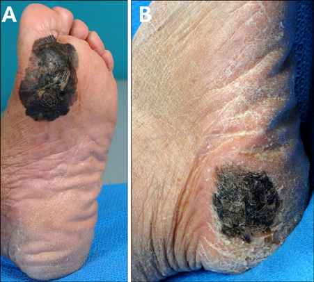
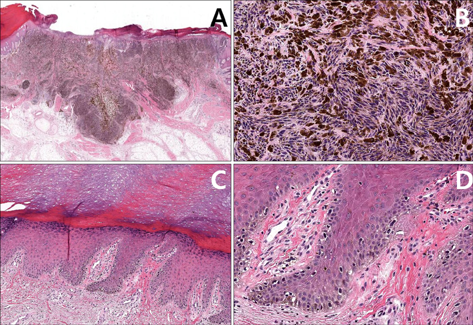
Double Primary Acral Lentiginous Melanoma of both Soles
- Affiliations
-
- 1Department of Dermatology, Samsung Medical Center, Sungkyunkwan University School of Medicine, Seoul, Korea. dylee@skku.edu
- KMID: 2368050
- DOI: http://doi.org/10.5021/ad.2017.29.1.129
Abstract
- No abstract available.
MeSH Terms
Figure
Reference
-
1. Savoia P, Quaglino P, Verrone A, Bernengo MG. Multiple primary melanomas: analysis of 49 cases. Melanoma Res. 1998; 8:361–366.
Article2. Hutcheson AC, McGowan JW 4th, Maize JC Jr, Cook J. Multiple primary acral melanomas in African-Americans: a case series and review of the literature. Dermatol Surg. 2007; 33:1–10.
Article3. Kim JY, Chi SG, Lee SJ, Kim HY, Lee WJ, Kim DW, et al. A case of multiple primary cutaneous melanoma. Korean J Dermatol. 2010; 48:435–439.4. Bae JM, Kim HO, Park YM. Progression from acral lentiginous melanoma in situ to invasive acral lentiginous melanoma. Ann Dermatol. 2009; 21:185–188.
Article5. Pollio A, Tomasi A, Pellacani G, Ruini C, Mandel VD, Fortuna G, et al. Multiple primary melanomas versus single melanoma of the head and neck: a comparison of genetic, diagnostic, and therapeutic implications. Melanoma Res. 2014; 24:267–272.
- Full Text Links
- Actions
-
Cited
- CITED
-
- Close
- Share
- Similar articles
-
- A Case of Thin Acral Lentiginous Melanoma with Lymph Node Metastasis and Regression
- Acral Lentiginous Melanoma Developing during Long-standing Atypical Melanosis: Usefulness of Dermoscopy for Detection of Early Acral Melanoma
- A Case of Acral Lentiginous Melanoma
- Acral Lentiginous Melanoma in situ
- Acral Lentiginous Melanoma, Indolent Subtype Diagnosed by En Bloc Excision: A Case Report


