Secondary Cutaneous Amyloidosis in a Patient with Mycosis Fungoides
- Affiliations
-
- 1Department of Dermatology, Dankook University Medical College, Cheonan, Korea. ivymyung@daum.net
- KMID: 2368032
- DOI: http://doi.org/10.5021/ad.2017.29.1.79
Abstract
- Secondary cutaneous amyloidosis refers to clinically unapparent amyloid deposits within the skin in association with a pre-existing skin condition or skin tumors, such as basal cell carcinoma, porokeratosis, solar elastosis, Bowen's disease, and mycosis fungoides. A 70-year-old woman presented with a 6-month history of asymptomatic multiple yellowish plaques on both legs. She had been diagnosed with mycosis fungoides 7 years ago and was treated with psoralen and ultraviolet A radiation (PUVA) therapy, narrow-band ultraviolet B (UVB) therapy, and acitretin for 5 years. Finally, she reached complete remission of mycosis fungoides. However, new yellowish lesions started to appear 1 year after discontinuing the phototherapy. A physical examination revealed multiple yellowish plaques on both extremities. The plaques were well circumscribed and slightly elevated. All laboratory tests were normal. A biopsy specimen showed multiple nodular deposits of eosinophilic amorphous material in papillary dermis and upper reticular dermis. The deposits represented apple green birefringence on Congo red stain viewed under polarized light. Acellular small nodules in the upper dermis consisted of randomly oriented, non-branching, 6.67~12.7 nm thick amyloid fibrils on electron microscopy. We report an interesting and rare case of secondary cutaneous amyloidosis after narrow-band UVB therapy and PUVA therapy in a patient with mycosis fungoides.
MeSH Terms
-
Acitretin
Aged
Amyloid
Amyloidosis*
Biopsy
Birefringence
Bowen's Disease
Carcinoma, Basal Cell
Congo Red
Dermis
Eosinophils
Extremities
Female
Ficusin
Humans
Leg
Microscopy, Electron
Mycosis Fungoides*
Phototherapy
Physical Examination
Plaque, Amyloid
Porokeratosis
PUVA Therapy
Skin
Ultraviolet Therapy
Acitretin
Amyloid
Congo Red
Ficusin
Figure
-

Fig. 1 (A~D) The patient was diagnosed with mycosis fungoides (MF) 7 years ago. Erythematous and brownish scaly oozing plaques on the entire body. MF stage: T3b-N0M0.
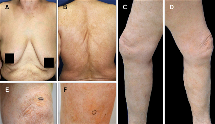
Fig. 2 (A, B) Complete remission of mycosis fungoides. The patient had been treated with psoralen and ultraviolet A radiation therapy, narrow-band phototherapy, and acitretin for 5 years and had reached complete remission of mycosis fungoides. (C~F) Multiple yellowish plaques on the both lower extremities including thigh, knee and lower leg. New yellowish lesions started to appear 1 year after discontinuing phototherapy. The plaques were well circumscribed and slightly elevated.

Fig. 3 (A) Multiple nodular deposits of fissured faintly eosinophilic amorphous material in the papillary dermis and upper reticular dermis (H&E, ×100). (B) Large, fissured, homogenous, eosinophilic masses in the papillary dermis (H&E, ×400). (C) The deposits were birefringence positive on Congo red staining viewed under polarized light microscopy (Congo red, ×100). (D) Electron micrographs show acellular small nodules consisting of randomly oriented, non-branching, non-anastomosing, 6.67~12.7 nm thick (mean, 9.3 nm) amyloid fibrils.
Reference
-
1. Breathnach SM. Amyloid and amyloidosis. J Am Acad Dermatol. 1988; 18:1–16.
Article2. Lee DY, Kim YJ, Lee JY, Kim MK, Yoon TY. Primary localized cutaneous nodular amyloidosis following local trauma. Ann Dermatol. 2011; 23:515–518.
Article3. Kumakiri M, Hashimoto K. Histogenesis of primary localized cutaneous amyloidosis: sequential change of epidermal keratinocytes to amyloid via filamentous degeneration. J Invest Dermatol. 1979; 73:150–162.
Article4. Li WM. Histopathology of primary cutaneous amyloidoses and systemic amyloidosis. Clin Dermatol. 1990; 8:30–35.
Article5. Hashimoto K, King LE Jr. Secondary localized cutaneous amyloidosis associated with actinic keratosis. J Invest Dermatol. 1973; 61:293–299.
Article6. Tsuji T, Asai Y, Hamada T. Secondary localized cutaneous amyloidosis in solar elastosis. Br J Dermatol. 1982; 106:469–475.
Article7. Aso M, Hagari Y, Nakamura K, Mihara M, Shimao S. A case of secondary cutaneous amyloidosis: epidermal keratinocytes produce amyloid in the cytoplasm. J Cutan Pathol. 1990; 17:176–181.
Article8. Holzmann H, Schott HJ. Amyloid demonstration in the skin in mycosis fungoides. Klin Wochenschr. 1965; 43:1061–1062.9. Schott HJ, Holzmann H. Detection of amyloid deposits in mycosis fungoides. Arch Klin Exp Dermatol. 1965; 222:632–641.10. Hashimoto K, Kumakiri M. Colloid-amyloid bodies in PUVA-treated human psoriatic patients. J Invest Dermatol. 1979; 72:70–80.
Article11. Greene I, Cox AJ. Amyloid deposition after psoriasis therapy with psoralen and long-wave ultraviolet light. Arch Dermatol. 1979; 115:1200–1202.
Article12. Powell AM, Albert S, Bhogal B, Black MM. Discoid lupus erythematosus with secondary amyloidosis. Br J Dermatol. 2005; 153:746–749.
Article13. Eto H, Hashimoto K, Kobayashi H, Fukaya T, Matsumoto M, Sun TT. Differential staining of cytoid bodies and skin-limited amyloids with monoclonal anti-keratin antibodies. Am J Pathol. 1984; 116:473–481.14. Zemheri IE, Ozkanli SS, Zindanci I, Senol S, Akbulak O, Topaloğlu Demir F. PUVA phototherapy-induced secondary amyloidosis in patients with mycosis fungoides: a rare adverse effect of phototherapy. Turk J Med Sci. 2014; 44:89–94.
Article15. Izumi K, Arita K, Horie K, Hoshina D, Shimizu H. Localized cutaneous amyloidosis associated with poikilodermatous mycosis fungoides. Acta Derm Venereol. 2014; 94:225–226.
Article
