Ann Dermatol.
2017 Feb;29(1):13-19. 10.5021/ad.2017.29.1.13.
Changing in the Epidemiology of Tinea Capitis among School Children in Egypt
- Affiliations
-
- 1Department of Medical Microbiology and Immunology, Faculty of Medicine, Fayoum University, Fayoum, Egypt. rhb00@fayoum.edu.eg
- 2Department of Public Health, Faculty of Medicine, Fayoum University, Fayoum, Egypt.
- 3Department of Dermatology & STD and Anderology, Faculty of Medicine, Fayoum University, Fayoum, Egypt.
- KMID: 2368022
- DOI: http://doi.org/10.5021/ad.2017.29.1.13
Abstract
- BACKGROUND
Tinea capitis remains a prevalent health problem among school-aged children.
OBJECTIVE
To estimate the prevalence of tinea capitis among primary school students, in Fayoum, Egypt with identification of etiological agents in both public and private primary schools.
METHODS
A cross-sectional study was conducted in twelve primary schools. The students were selected from different grades with a total number of 12,128 students. Hair and scalp were clinically examined for any lesions that may suspect tinea capitis and mycological samples were collected for direct microscopy and culture.
RESULTS
The prevalence of tinea capitis in the study group was 0.4% and higher in public than private schools (73.5% versus 26.5% respectively). Boys were more affected than girls with boy to girls' ratio 5:1. Intrafamily history of infection was present in 40.8% of tested group while 51% showed low social standard profile. Mycological culture revealed that Microsporum canis was the predominant isolated organism followed by M. audouinii (52% and 36% respectively).
CONCLUSION
M. canis is replacing Trichophyton violaceum as an etiology for tinea capitis in Egypt with lower prevalence rate than reported previously.
MeSH Terms
Figure
-

Fig. 1 The different types detected of the tinea capitis lesions. (A) Scaly type. (B) Black dot type. (C) Scaly & black dot type.
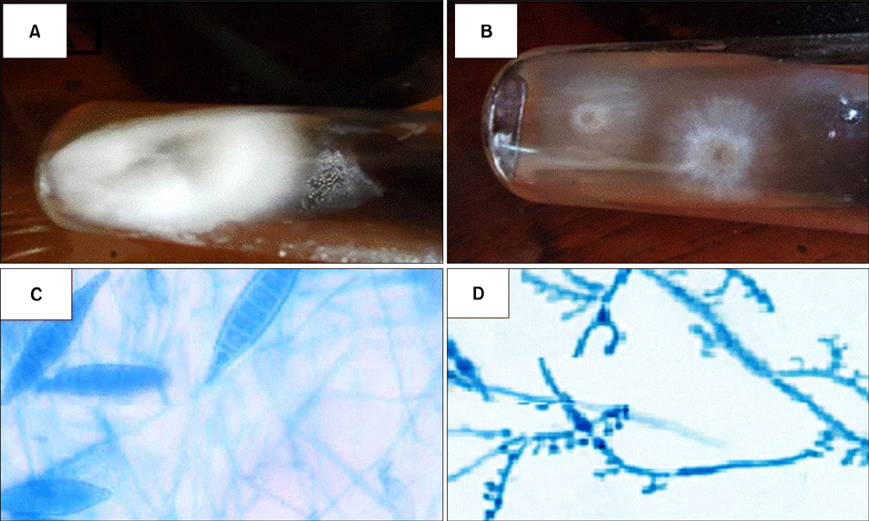
Fig. 2 Isolated dermatophytes. (A) Microsporum canis on Sabouraud dextrose agar (SDA). (B) M. audouinii on SDA. (C) Microscopic picture showing macroconidia of M. canis. (D) Microscopic picture showing pectinate bodies of M. audouinii.
Reference
-
1. Pérez-González M, Torres-Rodríguez JM, Martínez-Roig A, Segura S, Griera G, Triviño L, et al. Prevalence of tinea pedis, tinea unguium of toenails and tinea capitis in school children from Barcelona. Rev Iberoam Micol. 2009; 26:228–232.
Article2. Ghannoum M, Isham N, Hajjeh R, Cano M, Al-Hasawi F, Yearick D, et al. Tinea capitis in Cleveland: survey of elementary school students. J Am Acad Dermatol. 2003; 48:189–193.
Article3. Andrews MD, Burns M. Common tinea infections in children. Am Fam Physician. 2008; 77:1415–1420.4. Sidat MM, Correia D, Buene TP. Tinea capitis among children at one suburban primary school in the city of Maputo, Mozambique. Rev Soc Bras Med Trop. 2007; 40:473–475.
Article5. Degreef H. Clinical forms of dermatophytosis (ringworm infection). Mycopathologia. 2008; 166:257–265.
Article6. Bassiri-Jahromi S, Khaksari AA. Epidemiological survey of dermatophytosis in Tehran, Iran, from 2000 to 2005. Indian J Dermatol Venereol Leprol. 2009; 75:142–147.
Article7. Grimalt R. A practical guide to scalp disorders. J Investig Dermatol Symp Proc. 2007; 12:10–14.
Article8. Park JE, Park K. Textbook of preventive and social medicine. 7th ed. Jabalpur: Banarsidas Bhanot;1979. p. 81.9. Rippon JW. Dermatophytosis and dermatomycoses. Medical mycology-The pathogenic fungi and the pathogenic actinomycetes. 3rd ed. Philadelphia: WB Saunders;1988. p. 169–275.10. Kechia FA, Kouoto EA, Nkoa T, Nweze EI, Fokoua DC, Fosso S, et al. Epidemiology of tinea capitis among school-age children in Meiganga, Cameroon. J Mycol Med. 2014; 24:129–134.
Article11. Triviño-Duran L, Torres-Rodriguez JM, Martinez-Roig A, Cortina C, Belver V, Perez-Gonzalez M, et al. Prevalence of tinea capitis and tinea pedis in Barcelona schoolchildren. Pediatr Infect Dis J. 2005; 24:137–141.
Article12. Omar AA. Ringworm of the scalp in primary-school children in Alexandria: infection and carriage. East Mediterr Health J. 2000; 6:961–967.
Article13. Azab MM, Mahmoud NF, Abd Allah S, Hosny AM, Shehata AS, Mohamed RW. Dermatophytes isolated from clinical samples of children suffering from tinea capitis in Ismailia, Egypt. Aust J Basic Appl Sci. 2012; 6:38–42.14. Vijayakumar R, Muthukumar C, Kumar T, Saravanamuthu R. Characterization of Malassezia furfur and its control by using plant extracts. Indian J Dermatol. 2006; 51:145–148.
Article15. Geweely NSI. Antifungal activity of ozonized olive oil (Oleozone). Int J Agri Biol. 2006; 8:670–675.16. Fathi HI, al-Samarai AG. Prevalence of tinea capitis among schoolchildren in Iraq. East Mediterr Health J. 2000; 6:128–137.
Article17. El-Khalawany M, Shaaban D, Hassan H, Abdalsalam F, Eassa B, Abdel Kader A, et al. A multicenter clinicomycological study evaluating the spectrum of adult tinea capitis in Egypt. Acta Dermatovenerol Alp Pannonica Adriat. 2013; 22:77–82.18. Chapman JC, Daniel CR 3rd, Daniel JG, Daniel MP, Sullivan S, Howell D, et al. Tinea capitis caused by dermatophytes: a 15-year retrospective study from a Mississippi Dermatology Clinic. Cutis. 2011; 88:230–233.19. Abdel Fattah A, El-Gothamy Z. Tinea capitis in Egypt. Mykosen. 1967; 10:189–194.
Article20. Taha M, Amer M, Salem A, el Harras M. The perfect state of Trichophyton violaceum. Int J Dermatol. 1994; 33:493–495.21. Othman T, Vacher C. Tinea of the scalp in Egypt. Bull Soc Pathol Exot Filiales. 1983; 76:126–128.22. Ali-Shtayeh MS, Yaish S, Jamous RM, Arda H, Husein EI. Updating the epidemiology of dermatophyte infections in Palestine with special reference to concomitant dermatophytosis. J Mycol Med. 2015; 25:116–122.
Article23. Razzaq Adel AA, Sultan AO, Basmiah AM, Aftab A, Nabel N. Prevalence of tinea capitis in southern Kuwait. Mycoses. 2007; 50:317–320.
Article24. Abanmi A, Bakheshwain S, El Khizzi N, Zouman AR, Hantirah S, Al Harthi F, et al. Characteristics of superficial fungal infections in the Riyadh region of Saudi Arabia. Int J Dermatol. 2008; 47:229–235.
Article25. Ginter-Hanselmayer G, Weger W, Ilkit M, Smolle J. Epidemiology of tinea capitis in Europe: current state and changing patterns. Mycoses. 2007; 50:Suppl 2. 6–13.
Article
