Spontaneous Perforation of Common Bile Duct: Abscess Formation Presenting as a Choledochal Cyst
- Affiliations
-
- 1Department of Radiology, CHA Bundang Medical Center, CHA University, Gyeonggi-do, Korea. choikim75@gmail.com
- 2Department of Surgery, CHA Bundang Medical Center, CHA University, Gyeonggi-do, Korea.
- 3Department of Internal Medicine, CHA Bundang Medical Center, CHA University, Gyeonggi-do, Korea.
- KMID: 2366408
- DOI: http://doi.org/10.13104/imri.2016.20.4.254
Abstract
- Spontaneous perforation of the bile duct without any traumatic or iatrogenic injury is extremely rare. We report a case of abscess formation related to spontaneous perforation of the common bile duct by a gallstone, mimicked a cholecochal cyst.
MeSH Terms
Figure
-

Fig. 1 (a) Axial portal phase contrast enhanced CT shows a cystic lesion with a thick wall in the portocaval space (arrows). An asterisk denotes the common bile duct (CBD). (b) Axial portal phase contrast enhanced CT shows a cystic lesion with a thick wall and internal calcified gallstones (arrowhead) in the portocaval space. An asterisk denotes the CBD. (c, d) Axial and coronal portal phase contrast enhanced CT shows communication between the cystic lesion and the CBD (curved arrows). An asterisk denotes the CBD. (e) Axial portal phase contrast enhanced CT shows a small calcified gallstone in the distal CBD (arrowhead).

Fig. 2 (a) Three dimensional MRCP shows mild displacement of the CBD (arrows). The right posterior intrahepatic duct (arrowhead) drains at the mid CBD and the cystic duct (curved arrow) drains at the right posterior intrahepatic duct. (b) A cystic lesion in the portocaval space demonstrates high signal intensity on diffusion weighted MRI (b factor = 800 s/mm2) (arrow). (c) A cystic lesion in the portocaval space demonstrates low signal intensity on the apparent diffusion coefficient map (arrow).
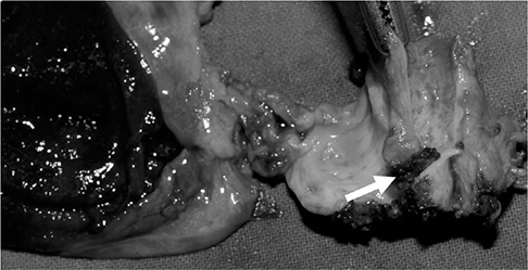
Fig. 3 The surgical specimen shows a mural defect in the CBD (arrow).
Reference
-
1. Kang SB, Han HS, Min SK, Lee HK. Nontraumatic perforation of the bile duct in adults. Arch Surg. 2004; 139:1083–1087.2. Wiseman K, Buczkowski AK, Chung SW, Francoeur J, Schaeffer D, Scudamore CH. Epidemiology, presentation, diagnosis, and outcomes of choledochal cysts in adults in an urban environment. Am J Surg. 2005; 189:527–531. discussion 531.3. Freeland J. Rupture of the hepatic duct. Lancet. 1882; 1:731–732.4. Mizutani S, Yagi A, Watanabe M, et al. T tube drainage for spontaneous perforation of the extrahepatic bile duct. Med Sci Monit. 2011; 17:CS8–CS11.5. Lee HK, Han HS, Lee JH, Min SK. Nontraumatic perforation of the bile duct treated with laparoscopic surgery. J Laparoendosc Adv Surg Tech A. 2005; 15:329–332.6. Piotrowski JJ, Van Stiegmann G, Liechty RD. Spontaneous bile duct rupture in pregnancy. HPB Surg. 1990; 2:205–209.7. Thompson CM, Saad NE, Quazi RR, Darcy MD, Picus DD, Menias CO. Management of iatrogenic bile duct injuries: role of the interventional radiologist. Radiographics. 2013; 33:117–134.8. Ouaissi M, Kianmanesh R, Belghiti J, et al. Todani Type II congenital bile duct cyst: European Multicenter Study of the French Surgical Association and literature review. Ann Surg. 2015; 262:130–138.9. Qayyum A. Diffusion-weighted imaging in the abdomen and pelvis: concepts and applications. Radiographics. 2009; 29:1797–1810.10. Megison SM, Votteler TP. Management of common bile duct obstruction associated with spontaneous perforation of the biliary tree. Surgery. 1992; 111:237–239.
