Anatomical variants of the superficial temporal artery in patients with microtia: a pilot descriptive study
- Affiliations
-
- 1Division of Plastic Surgery, Department of Surgery, University Kebangsaan Malaysia Medical Centre, Kuala Lumpur, Malaysia. farrahhani@gmail.com
- 2Department of General Surgery, Queen Elizabeth Hospital, Kota Kinabalu, Malaysia.
- 3Department of Anatomy, Universiti Kebangsaan Malaysia Medical Centre, Kuala Lumpur, Malaysia.
- 4Prince Court Medical Centre, Kuala Lumpur, Malaysia.
- KMID: 2365557
- DOI: http://doi.org/10.5115/acb.2016.49.4.273
Abstract
- Superficial temporal artery (STA) based pedicled fascial flap plays a pivotal role in ear reconstruction for microtia patients. There is paucity of literature on the anatomy of the STA in microtia patients. The present study aimed to describe any possible anatomical variations seen in the STA of patients afflicted with microtia. Pre-operative carotid computer tomographic angiography images of patients under the microtia database of Plastic and Reconstructive Surgery Unit at a tertiary medical centre were selected and 3-dimensionally reconstructed. Measurements were made on the 3D reconstructed computed tomographic angiography images of the STA on both the sides of the microtic ear and the non-microtic ear to assess its various anatomical parameters. We managed to obtain a total of 39 computed tomographic angiography images of STAs for analysis. There was a significant difference in the number of main branches of STA between the two groups (P=0.006). The proportion of ears with 2 main branches was higher in the non-microtia group (89.5%) compared to the microtia group (45.0%). A significant difference was found in the STA diameter between the two groups (P=0.012). The mean diameter of STA in the non-microtia group was larger by 0.4 mm. Furthermore, the median angle of STA was larger on the side of the non-microtic ears compared to that of microtic ears by 24.5°, with a P-value of 0.011. The results of the study may be of clinical importance while planning and performing ear reconstructive surgeries using STA based pedicled fascial flaps.
Keyword
Figure
-
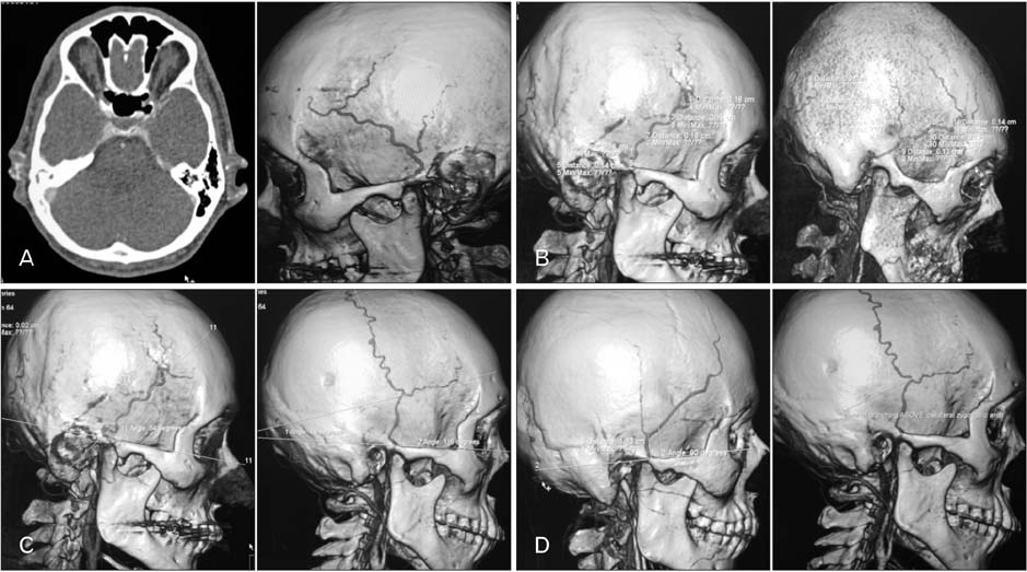
Fig. 1 (A) 3D reconstruction of external carotid computed tomographic angiography. (B) Measurement of the diameter of superficial temporal artery (STA) and its main branches. (C) Measurement of the angle of STA and its main branches in relation to a plane drawn parallel to the upper border of the ipsilateral zygomatic arch. (D) Measurement of the distance and level of STA branching point in relation to the upper border of the ipsilateral zygomatic arch.
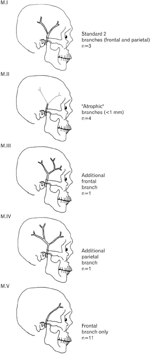
Fig. 2 Anatomical variations of the superficial temporal artery in microtia subjects.
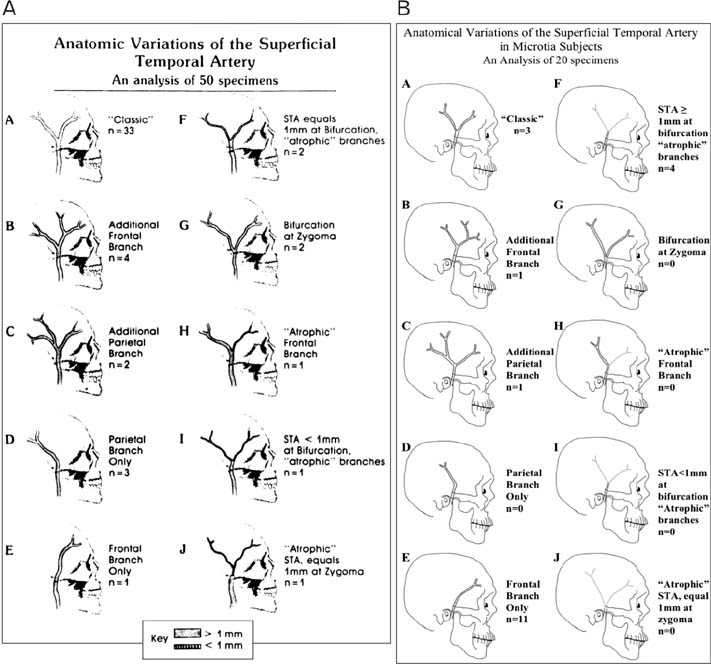
Fig. 3 (A) Classification of superficial temporal artery (STA) by anatomic variations in normal population, according to Marano et al. [4]. Adapted from Marano et al. Neurosurgery 1985;16:786-90 [4], with permission from Wolters Kluwer. (B) Preponderance of different anatomical variants of STA in the microtia population compared to that described by Marano et al. [4] in the normal population. Note that the main variants found in this study is the type “E” variants, i.e., STA with frontal branch only. Also, it was noted that the mean diameters of the STA and its branches were smaller in the microtia population compared to the normal population described by Marano et al. [4].
Reference
-
1. Gardner ED, Gray DJ, O'Rahilly R. Anatomy: a regional study of human structure. 4th ed. Philadelphia, PA: W.B. Saunders;1975.2. Abul-Hassan HS, von Drasek Ascher G, Acland RD. Surgical anatomy and blood supply of the fascial layers of the temporal region. Plast Reconstr Surg. 1986; 77:17–28.3. Stock AL, Collins HP, Davidson TM. Anatomy of the superficial temporal artery. Head Neck Surg. 1980; 2:466–469.4. Marano SR, Fischer DW, Gaines C, Sonntag VK. Anatomical study of the superficial temporal artery. Neurosurgery. 1985; 16:786–790.5. Chen TH, Chen CH, Shyu JF, Wu CW, Lui WY, Liu JC. Distribution of the superficial temporal artery in the Chinese adult. Plast Reconstr Surg. 1999; 104:1276–1279.6. Yamada A, Ueda K, Yorozuya-Shibazaki R. External ear reconstruction in hemifacial microsomia. J Craniofac Surg. 2009; 20:Suppl 2. 1787–1793.7. Melnick M, Myrianthopoulos NC, Paul NW. External ear malformations: epidemiology, genetics, and natural history. Birth Defects Orig Artic Ser. 1979; 15:i–ix. 1–140.8. Kelley PE, Scholes MA. Microtia and congenital aural atresia. Otolaryngol Clin North Am. 2007; 40:61–80.9. Brent B. The correction of mi-rotia with autogenous cartilage grafts: I. The classic deformity? Plast Reconstr Surg. 1980; 66:1–12.10. Nagata S. A new method of total reconstruction of the auricle for microtia. Plast Reconstr Surg. 1993; 92:187–201.11. Tanzer RC. Total reconstruction of the auricle. The evolution of a plan of treatment. Plast Reconstr Surg. 1971; 47:523–533.12. Brent B. The correction of microtia with autogenous cartilage grafts: II. Atypical and complex deformities. Plast Reconstr Surg. 1980; 66:13–21.13. Park C, Lew DH, Yoo WM. An analysis of 123 temporoparietal fascial flaps: anatomic and clinical considerations in total auricular reconstruction. Plast Reconstr Surg. 1999; 104:1295–1306.14. Pinar YA, Govsa F. Anatomy of the superficial temporal artery and its branches: its importance for surgery. Surg Radiol Anat. 2006; 28:248–253.15. Tayfur V, Edizer M, Magden O. Anatomic bases of superficial temporal artery and temporal branch of facial nerve. J Craniofac Surg. 2010; 21:1945–1947.16. Kim BS, Jung YJ, Chang CH, Choi BY. The anatomy of the superficial temporal artery in adult koreans using 3-dimensional computed tomographic angiogram: clinical research. J Cerebrovasc Endovasc Neurosurg. 2013; 15:145–151.
- Full Text Links
-
- Actions
-
Cited
- CITED
-
- Close
- Share
-
- Similar articles
-
- Aneurysms of the superficial temporal artery
- A Case Report of Posttraumatic Pseudoaneurysm of the Superficial Temporal Artery
- Traumatic Pseudoaneurysm of the Superficial Temporal Artery
- Topographical Relationship of the Superficial Temporal Vessels and the Auriculotemporal Nerve
- Ear Elevation using the Superficial Temporal Fascia Harvested Through Minimal Incision
