Adrenal Castleman's Disease Mimicking Other Adrenal Neoplasms: A Case Report
- Affiliations
-
- 1Department of Radiology, Pusan National University Hospital, Pusan National University School of Medicine, Busan, Korea. leenk77@hanmail.net
- 2Department of Urology, Pusan National University Hospital, Pusan National University School of Medicine, Busan, Korea.
- 3Department of Pathology, Pusan National University Hospital, Pusan National University School of Medicine, Busan, Korea.
- KMID: 2365055
- DOI: http://doi.org/10.3348/jksr.2017.76.1.73
Abstract
- We present a rare case of adrenal Castleman's disease with hyaline vascular type mimicking other adrenal neoplasms in a 65-year-old woman. Although rare, the hyaline vascular type of adrenal Castleman's disease should be included in the differential diagnosis if an adrenal mass shows a well-defined, highly enhancing solid adrenal mass with peripheral rim enhancement, multiple satellite lymph nodes, and peritoneal thickening around the dominant mass on computed tomography as shown in this patient.
MeSH Terms
Figure
-

Fig. 1 Adrenal Castleman's disease mimicking other adrenal neoplasms in a 65-year-old woman. A. Pre-enhanced CT showing a well-defined homogenous soft tissue density mass of 5.0 cm in long dimension (arrow) in the left adrenal gland. B. Contrast-enhanced CT showing a highly enhancing mass with peripheral rim enhancement (arrow). Peritoneal thickening (arrowhead) surrounding the mass is noted. C. Contrast-enhanced CT showing multiple small enhancing retroperitoneal lymph nodes (arrow). Increased fat stranding in the left perirenal space was also noted.
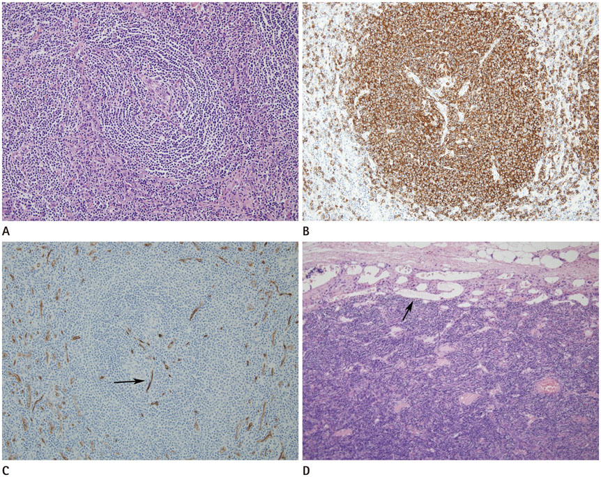
Fig. 2 Hyaline-vascular type Castleman's disease of the adrenal gland in a 65-year-old woman. A. Photomicrograph (original magnification, × 200 hematoxylin-eosin stain) of the mass showing lymphoid follicles with small hyalinized germinal centers and broad mantle zone. Mantle zone lymphocytes are arranged in concentric rings (“onion skin” pattern). B. The “onion skin” pattern is highlighted by immunochemical staining for CD20 (original magnification, × 200). C. Photomicrograph (original magnification, × 200; immunochemical staining for CD34) of the mass showing some follicles with penetration by blood vessels (“lollipop follicle”, arrow). D. Photomicrograph (original magnification, × 100 hematoxylin-eosin stain) of the mass showing prominent vascular proliferation (arrow) in the periphery of the mass.
Reference
-
1. Shringarpure S, Sivaraman PB, Parmeswaran A. Castleman’s disease: a rare differential diagnosis for retroperitoneal tumors. Urol Ann. 2010; 2:44–45.2. Keller AR, Hochholzer L, Castleman B. Hyaline-vascular and plasma-cell types of giant lymph node hyperplasia of the mediastinum and other locations. Cancer. 1972; 29:670–683.3. Cronin DM, Warnke RA. Castleman disease: an update on classification and the spectrum of associated lesions. Adv Anat Pathol. 2009; 16:236–246.4. Ko HS, Woo JY, Hong HS, Jung AY, Yang I, Lee Y. Castleman disease in the kidney and retroperitoneum mimicking renal cell carcinoma with retroperitoneal lymphadenopathy: a case report. J Korean Soc Radiol. 2012; 67:397–400.5. Bonekamp D, Horton KM, Hruban RH, Fishman EK. Castleman disease: the great mimic. Radiographics. 2011; 31:1793–1807.6. Müssig K, Horger M, Wehrmann M. Adrenal Castleman’s disease. Ann Hematol. 2007; 86:63–65.7. Meador TL, McLarney JK. CT features of Castleman disease of the abdomen and pelvis. AJR Am J Roentgenol. 2000; 175:115–118.8. Zhou LP, Zhang B, Peng WJ, Yang WT, Guan YB, Zhou KR. Imaging findings of Castleman disease of the abdomen and pelvis. Abdom Imaging. 2008; 33:482–488.9. Zheng X, Pan K, Cheng J, Dong L, Yang K, Wu E. Localized Castleman disease in retroperitoneum: newly discovered features by multi-detector helical CT. Abdom Imaging. 2008; 33:489–492.10. Johnson PT, Horton KM, Fishman EK. Adrenal mass imaging with multidetector CT: pathologic conditions, pearls, and pitfalls. Radiographics. 2009; 29:1333–1351.
- Full Text Links
-
- Actions
-
Cited
- CITED
-
- Close
- Share
-
- Similar articles
-
- Pyogenic Adrenal Cyst in Newborn
- A case of primary bilateral adrenal lymphoma with partial adrenal insufficiency
- Adrenal Tuberculosis Mimicking a Malignant Tumor with Primary Adrenal Insufficiency
- A Case of Adrenal Tuberculosis Combined with Tuberculous Peritonitis-Induced Adrenal Crisis
- A Case of an Adrenal Hemangioma Mimicking a Pancreatic Tail Tumor
