J Korean Soc Surg Hand.
2016 Dec;21(4):198-204. 10.12790/jkssh.2016.21.4.198.
Biomechanical Analysis of Resorbable Barbed Suture Tenorrhaphy
- Affiliations
-
- 1Department of Plastic and Reconstructive Surgery, Soonchunhyang University College of Medicine, Bucheon, Korea.
- 2Jayjun Plastic Surgery and Aesthetic, Seoul, Korea.
- 3Department of Plastic and Reconstructive Surgery, National Medical Center, Seoul, Korea. sangwind@hanmail.net
- KMID: 2364210
- DOI: http://doi.org/10.12790/jkssh.2016.21.4.198
Abstract
- PURPOSE
To evaluate the tensile strength and repair-site profile of a technique of resorbable barbed suture tenorrhaphy.
METHODS
Forty-eight flexor digitorum profundus tendons were collected from the 8 adult cadavers. In the test group, the tendons were sutured using absorbent 2-0 barb knotless sutures in a 2-strand or 4-strand zig-zag pattern. In the control group, 2-0 Prolene and 3-0 polydioxanone (PDS) were used to suture the tendons using the 2-stand Modified Kessler method and the 4-strand cruciate suture method. Using a tensile force measurement machine, the breaking load (N) and the stiffness (N/mm) were measured. The types of rupture were categorized into suture breaking, knot rupture, and pullout.
RESULTS
In the comparative analysis between the absorbent 2-0 Quill (Angiotech Pharmaceuticals, Canada) suture that used the 2-strand core suture and the 3-0 PDS and 2-0 Prolene sutures, the average breaking load for the 2-0 Quill suture was 26.83±7.47 N, and 21.96±6.78 N and 17.20±4.93 N for the 2-0 Prolene and 3-0 PDS sutures. In the comparison using the 4-strand core suture, the average breaking load for the 2-0 Quill suture was 62.50±13.34 N, and 22.35±5.72 N and 18.67±4.27 N for the 2-0 prolene and 3-0 PDS sutures. The most common type of rupture were knot rupture.
CONCLUSION
For flexor tendon sutures using the absorbent barb sutures, compared to the conventional 2-0 Prolene or 3-0 PDS sutures, absorbent barbed sutures have a higher tensile strength.
Keyword
MeSH Terms
Figure
-

Fig. 1. Test machine for the (A) wire strength test and (B) suture strength test.
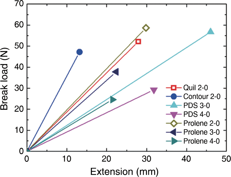
Fig. 2. Graph of break load and extension. PDS, polydioxanone.
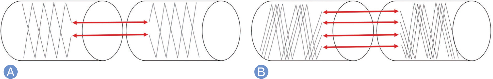
Fig. 3. (A) 2-strand and (B) 4-strand barbed suture technique.

Fig. 4. Analysis of results. The panel shows (A) suture break, (B) knot rupture, and (C) pullout, respectively.

Fig. 5. Load of failure in 2-strand tenorrhaphy. NS, not significant; PDS, polydioxanone. A p<0.05 was considered significant.
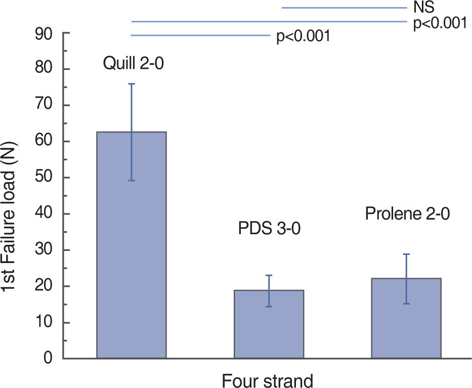
Fig. 6. Load of failure in 4-strand tenorrhaphy. S, not significant; PDS, polydioxanone. A p<0.05 was considered significant.
Reference
-
1. Strickland JW. Development of flexor tendon surgery: twenty-five years of progress. J Hand Surg Am. 2000; 25:214–35.
Article2. Aoki M, Kubota H, Pruitt DL, Manske PR. Biomechanical and histologic characteristics of canine flexor tendon repair using early postoperative mobilization. J Hand Surg Am. 1997; 22:107–14.
Article3. Bainbridge LC, Robertson C, Gillies D, Elliot D. A comparison of post-operative mobilization of flexor tendon repairs with “passive flexion-active extension” and “controlled active motion” techniques. J Hand Surg Br. 1994; 19:517–21.
Article4. Silfverskiold KL, May EJ. Flexor tendon repair in zone II with a new suture technique and an early mobilization program combining passive and active flexion. J Hand Surg Am. 1994; 19:53–60.5. Sirotakova M, Elliot D. Early active mobilization of primary repairs of the flexor pollicis longus tendon with two Kessler two-strand core sutures and a strengthened circumferential suture. J Hand Surg Br. 2004; 29:531–5.
Article6. Ketchum LD. Suture materials and suture techniques used in tendon repair. Hand Clin. 1985; 1:43–53.
Article7. Bunnell S. Gig pull-out suture for tendons. J Bone Joint Surg Am. 1954; 36:850–1.
Article8. Jennings ER, Mansberger AR Jr, Smith EP Jr, Yeager GH. A new technique in primary tendon repair. Surg Gynecol Obstet. 1952; 95:597–600.9. Parikh PM, Davison SP, Higgins JP. Barbed suture tenorrhaphy: an ex vivo biomechanical analysis. Plast Reconstr Surg. 2009; 124:1551–8.
Article10. Trail IA, Powell ES, Noble J. An evaluation of suture materials used in tendon surgery. J Hand Surg Br. 1989; 14:422–7.
Article11. Momose T, Amadio PC, Zhao C, Zobitz ME, An KN. The effect of knot location, suture material, and suture size on the gliding resistance of flexor tendons. J Biomed Mater Res. 2000; 53:806–11.
Article12. McKenzie AR. An experimental multiple barbed suture for the long flexor tendons of the palm and fingers: preliminary report. J Bone Joint Surg Br. 1967; 49:440–7.13. Amadio P, An KN, Ejeskar A, et al. IFSSH flexor tendon committee report. J Hand Surg Br. 2005; 30:100–16.14. Clemente A, Bergamin F, Surace C, Lepore E, Pugno N. Barbed suture vs conventional tenorrhaphy: biomechanical analysis in an animal model. J Orthop Traumatol. 2015; 16:251–7.
Article
- Full Text Links
-
- Actions
-
Cited
- CITED
-
- Close
- Share
-
- Similar articles
-
- Absorbable Barbed Suture for the Repair of Tendo Calcaneus in Rabbits
- Barbed sutures versus conventional tenorrhaphy in flexor tendon repair: An ex vivo biomechanical analysis
- Postoperative mechanical small bowel obstruction induced by V-Loc barbed absorbable suture after laparoscopic distal gastrectomy
- Surgical and Clinical Outcomes Associated With the Use of Barbed Sutures and Self-Adhering Mesh System and Polymeric Glue for Wound Closure in Multilevel or Revision Spinal Surgery: A Matched Cohort Comparative Study With Conventional Wound Closure Procedure
- Biomechanical Analysis of Tendon Suture Tecniques
