J Korean Assoc Oral Maxillofac Surg.
2015 Aug;41(4):190-193. 10.5125/jkaoms.2015.41.4.190.
A comparative analysis of patients with mesiodenses: a clinical and radiological study
- Affiliations
-
- 1Department of Oral and Maxillofacial Surgery, School of Dentistry, Chosun University, Gwangju, Korea. sgckim@chosun.ac.kr
- 2Department of Oral and Maxillofacial Surgery, Section of Dentistry, Seoul National University Bundang Hospital, Seongnam, Korea.
- 3Department of Pediatric Dentistry, School of Dentistry, Chosun University, Gwangju, Korea.
- KMID: 2364015
- DOI: http://doi.org/10.5125/jkaoms.2015.41.4.190
Abstract
OBJECTIVES
A mesiodens appears most commonly as a supernumerary tooth impacted in the anterior maxilla. The purpose of this study is analyze mesiodens clinically.
MATERIALS AND METHODS
Gender, crown form, direction of impaction, relation to permanent incisors, and chief complaints of patients with extracted mesiodens were analyzed.
RESULTS
Patients were analyzed for motivation to visit the hospital; 85.4% of the patients were referred from other hospitals. Mesiodens was more common in males than in females (3.7:1), and 70.1% of patients had only one mesiodens, while 29.6% had two mesiodenses. Of the mesiodenses, 61.4% were of the aconical form, and the most common direction was upward (62.4%), followed by the normal position (26.0%) and the horizontal position (11.6%). The mesiodenses caused orthodontic problems with the permanent incisors in 46.3% of cases. Mesiodens associated with dentigerous cyst was rarely observed in our patient group.
CONCLUSION
Mesiodens is more common in males than in females and often affects the permanent incisors. Thus, careful clinical and radiological evaluations of mesiodenses are important.
Keyword
MeSH Terms
Figure
-
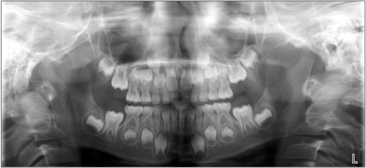
Fig. 1 A mesiodens found in an oral panoramic X-ray.
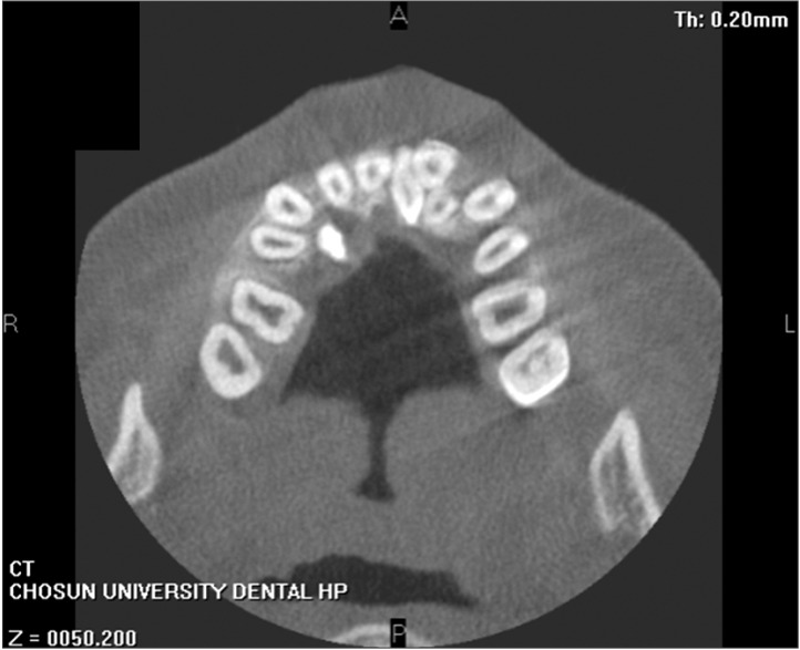
Fig. 2 An impacted conical-shaped mesiodens.
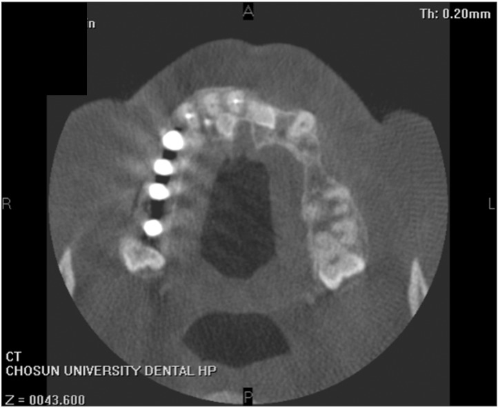
Fig. 3 Two horizontally impacted mesiodenses.
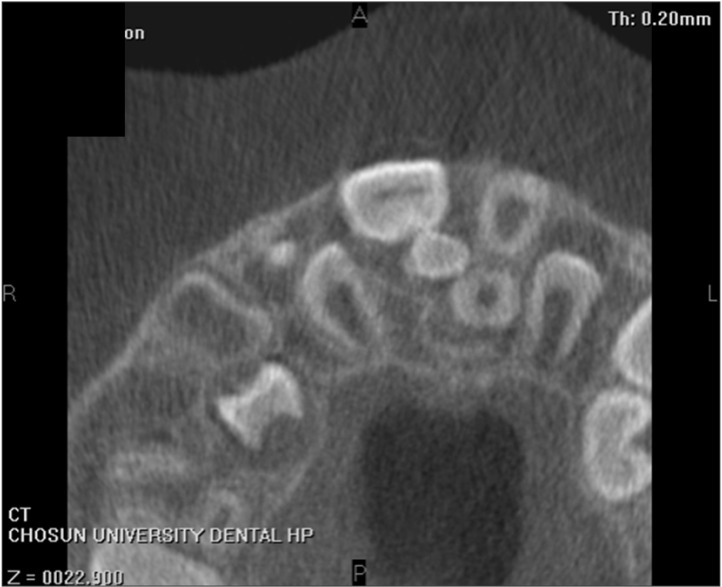
Fig. 4 A mesiodens impacted on the palatal side of the central incisor.
Fig. 5 A mesiodens impacted in the opposite direction.
Reference
-
1. von Arx T. Anterior maxillary supernumerary teeth: a clinical and radiographic study. Aust Dent J. 1992; 37:189–195. PMID: 1627067.2. Lustmann J, Bodner L. Dentigerous cysts associated with supernumerary teeth. Int J Oral Maxillofac Surg. 1988; 17:100–102. PMID: 3133415.
Article3. Dinkar AD, Dawasaz AA, Shenoy S. Dentigerous cyst associated with multiple mesiodens: a case report. J Indian Soc Pedod Prev Dent. 2007; 25:56–59. PMID: 17456972.
Article4. Kim SG, Lee SH. Mesiodens: a clinical and radiographic study. J Dent Child (Chic). 2003; 70:58–60. PMID: 12762611.5. Orhan AI, Ozer L, Orhan K. Familial occurrence of nonsyndromal multiple supernumerary teeth. A rare condition. Angle Orthod. 2006; 76:891–897. PMID: 17029528.6. Ray D, Bhattacharya B, Sarkar S, Das G. Erupted maxillary conical mesiodens in deciduous dentition in a Bengali girl--a case report. J Indian Soc Pedod Prev Dent. 2005; 23:153–155. PMID: 16224138.7. Rajab LD, Hamdan MA. Supernumerary teeth: review of the literature and a survey of 152 cases. Int J Paediatr Dent. 2002; 12:244–254. PMID: 12121534.
Article8. Gallas MM, García A. Retention of permanent incisors by mesiodens: a family affair. Br Dent J. 2000; 188:63–64. PMID: 10689766.
Article9. Fernández Montenegro P, Valmaseda Castellón E, Berini Aytés L, Gay Escoda C. Retrospective study of 145 supernumerary teeth. Med Oral Patol Oral Cir Bucal. 2006; 11:E339–E344. PMID: 16816819.10. Meighani G, Pakdaman A. Diagnosis and management of supernumerary (mesiodens): a review of the literature. J Dent (Tehran). 2010; 7:41–49. PMID: 21998774.
- Full Text Links
-
- Actions
-
Cited
- CITED
-
- Close
- Share
-
- Similar articles
-
- Three dimensional evaluation of impacted mesiodens using dental cone beam CT
- Clinical comparative Study Between Uncemented Anatomic Stem with and Without Lateral Porous Pad
- A comparative study on radiological and endoscopic examinations of the stomach cancer
- Assessment of Clinical and Radiological Parameters in Spinal Tuberculosis: Comparison between Human Immunodeficiency Virus-Positive and Human Immunodeficiency Virus-Negative Patients
- Comparative Analysis between Patellar Resurfacing and Retention in Total Knee Arthroplasty: 5-year Follow-up Result
