Comparative study on the osseointegration of implants in dog mandibles according to the implant surface treatment
- Affiliations
-
- 1Department of Oral and Maxillofacial Surgery, School of Dentistry, Chosun University, Gwangju, Korea. sgckim@chosun.ac.kr
- 2Department of Oral and Maxillofacial Surgery, Section of Dentistry, Konyang University Hospital, Daejeon, Korea.
- 3Department of Pathology, School of Medicine, Chosun University, Gwangju, Korea.
- 4Department of Dental Hygiene, Kangwon National University, Samcheok, Korea.
- KMID: 2364001
- DOI: http://doi.org/10.5125/jkaoms.2016.42.6.345
Abstract
OBJECTIVES
This study compared the impact of implant surface treatment on the stability and osseointegration of implants in dog mandibles.
MATERIALS AND METHODS
Six adult dogs received a total of 48 implants that were prepared using four different surface treatments; resorbable blast media (RBM), hydroxyapatite (HA), hydrothermal-treated HA, and sand blasting and acid etching (SLA). Implants were installed, and dogs were separated into 2- and 4-week groups. Implant stability was evaluated via Periotest M, Osstell Mentor, and removal torque analyzers. A histomorphometric analysis was also performed.
RESULTS
The stability evaluation showed that all groups generally had satisfactory values. The histomorphometric evaluation via a light microscope revealed that the HA surface implant group had the highest ratio of new bone formation on the entire fixture. The hydrothermal-treated HA surface implant group showed a high ratio of bone-to-implant contact in the upper half of the implant area.
CONCLUSION
The hydrothermal-treated HA implant improved the bone-to-implant contact ratio on the upper fixture, which increased the implant stability.
Keyword
MeSH Terms
Figure
-
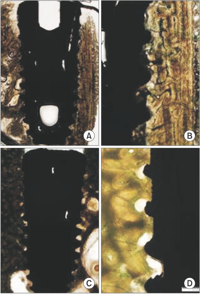
Fig. 1 Resorbable blast media surface fixture light micrographs in the 2-week (A, B) and 4-week (C, D) groups. The new bone formation area and bone-implant contact increased in the 4-week group (Villanueva osteochrome bone stain, A, C: ×12.5, B, D: ×40).
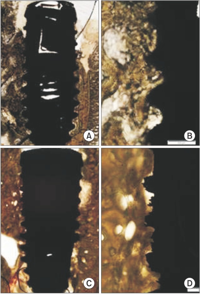
Fig. 2 Hydroxyapatite (HA) surface fixture light micrographs in the 2-week (A, B) and 4-week (C, D) groups. The new bone formation area (NBFA) and bone-implant contact increased in the 4-week group. Compact new bone formation was identified around the implant. The best NBFA value was obtained in this HA group (Villanueva osteochrome bone stain, A, C: ×12.5, B, D: ×40).
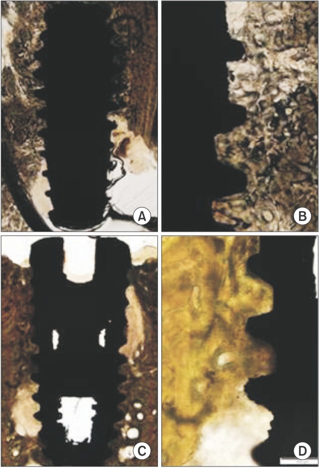
Fig. 3 Hydrothermal-treated hydroxyapatite (HA) surface fixture light micrographs in the 2-week (A, B) and 4-week (C, D) groups. The new bone formation area and bone-implant contact (BIC) increased in the 4-week group. Continuous BIC was identified along the surface of the implant. The best BIC value was obtained in this hydrothermal-treated HA group (Villanueva osteochrome bone stain, A, C: ×12.5, B, D: ×40).
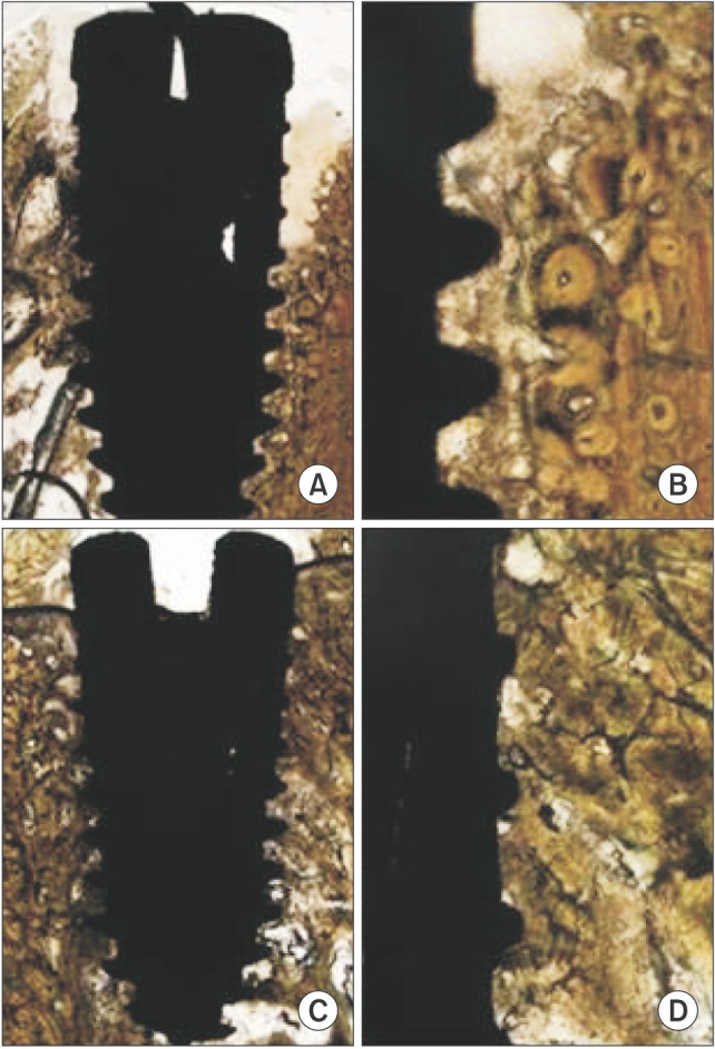
Fig. 4 The sand blasting and acid etching surface fixture light micrographs in the 2-week (A, B) and 4-week (C, D) groups. The new bone formation area and bone-implant contact increased in the 4-week group (Villanueva osteochrome bone stain, A, C: ×12.5, B, D: ×40).
Reference
-
1. Mueller CK, Thorwarth M, Schmidt M, Schlegel KA, Schultze-Mosgau S. Comparative analysis of osseointegration of titanium implants with acid-etched surfaces and different biomolecular coatings. Oral Surg Oral Med Oral Pathol Oral Radiol Endod. 2011; 112:726–736. PMID: 21441047.
Article2. Abuhussein H, Pagni G, Rebaudi A, Wang HL. The effect of thread pattern upon implant osseointegration. Clin Oral Implants Res. 2010; 21:129–136. PMID: 19709058.
Article3. de Vicente JC, Recio O, Martín-Villa L, Junquera LM, López-Arranz JS. Histomorphometric evaluation of guided bone regeneration around implants with SLA surface: an experimental study in beagle dogs. Int J Oral Maxillofac Surg. 2006; 35:1047–1053. PMID: 16973332.
Article4. Albrektsson T, Brånemark PI, Hansson HA, Lindström J. Osseointegrated titanium implants. Requirements for ensuring a long-lasting, direct bone-to-implant anchorage in man. Acta Orthop Scand. 1981; 52:155–170. PMID: 7246093.5. Brånemark PI, Adell R, Breine U, Hansson BO, Lindström J, Ohlsson A. Intra-osseous anchorage of dental prostheses. I. Experimental studies. Scand J Plast Reconstr Surg. 1969; 3:81–100. PMID: 4924041.6. Shalabi MM, Gortemaker A, Van't Hof MA, Jansen JA, Creugers NH. Implant surface roughness and bone healing: a systematic review. J Dent Res. 2006; 85:496–500. PMID: 16723643.
Article7. Thompson JI, Gregson PJ, Revell PA. Analysis of push-out test data based on interfacial fracture energy. J Mater Sci Mater Med. 1999; 10:863–868. PMID: 15347966.8. Buser D, Schenk RK, Steinemann S, Fiorellini JP, Fox CH, Stich H. Influence of surface characteristics on bone integration of titanium implants. A histomorphometric study in miniature pigs. J Biomed Mater Res. 1991; 25:889–902. PMID: 1918105.
Article9. Le Guéhennec L, Soueidan A, Layrolle P, Amouriq Y. Surface treatments of titanium dental implants for rapid osseointegration. Dent Mater. 2007; 23:844–854. PMID: 16904738.
Article10. Buser D, Broggini N, Wieland M, Schenk RK, Denzer AJ, Cochran DL, et al. Enhanced bone apposition to a chemically modified SLA titanium surface. J Dent Res. 2004; 83:529–533. PMID: 15218041.
Article11. Ellingsen JE, Johansson CB, Wennerberg A, Holmén A. Improved retention and bone-tolmplant contact with fluoride-modified titanium implants. Int J Oral Maxillofac Implants. 2004; 19:659–666. PMID: 15508981.12. Jeong R, Marin C, Granato R, Suzuki M, Gil JN, Granjeiro JM, et al. Early bone healing around implant surfaces treated with variations in the resorbable blasting media method. A study in rabbits. Med Oral Patol Oral Cir Bucal. 2010; 15:e119–e125. PMID: 19767688.
Article13. Meredith N, Book K, Friberg B, Jemt T, Sennerby L. Resonance frequency measurements of implant stability in vivo. A cross-sectional and longitudinal study of resonance frequency measurements on implants in the edentulous and partially dentate maxilla. Clin Oral Implants Res. 1997; 8:226–233. PMID: 9586467.14. Ratner BD, Porter SC. Surfaces in biology and biomaterials: description and characterization. In : Brash JL, Wojciechowski PW, editors. Interfacial phenomena and bioproducts. New York: Marcel Dekker;1996. p. 57–83.15. Davies JE. Understanding peri-implant endosseous healing. J Dent Educ. 2003; 67:932–949. PMID: 12959168.
Article16. Schwarz ML, Kowarsch M, Rose S, Becker K, Lenz T, Jani L. Effect of surface roughness, porosity, and a resorbable calcium phosphate coating on osseointegration of titanium in a minipig model. J Biomed Mater Res A. 2009; 89:667–678. PMID: 18442101.
Article17. Olivé J, Aparicio C. Periotest method as a measure of osseointegrated oral implant stability. Int J Oral Maxillofac Implants. 1990; 5:390–400. PMID: 2094658.18. Coelho PG, Granjeiro JM, Romanos GE, Suzuki M, Silva NR, Cardaropoli G, et al. Basic research methods and current trends of dental implant surfaces. J Biomed Mater Res B Appl Biomater. 2009; 88:579–596. PMID: 18973274.
Article19. Piattelli M, Scarano A, Paolantonio M, Iezzi G, Petrone G, Piattelli A. Bone response to machined and resorbable blast material titanium implants: an experimental study in rabbits. J Oral Implantol. 2002; 28:2–8. PMID: 12498456.
Article20. Granato R, Marin C, Suzuki M, Gil JN, Janal MN, Coelho PG. Biomechanical and histomorphometric evaluation of a thin ion beam bioceramic deposition on plateau root form implants: an experimental study in dogs. J Biomed Mater Res B Appl Biomater. 2009; 90:396–403. PMID: 19107801.
Article21. Yang GL, He FM, Yang XF, Wang XX, Zhao SF. Bone responses to titanium implants surface-roughened by sandblasted and double etched treatments in a rabbit model. Oral Surg Oral Med Oral Pathol Oral Radiol Endod. 2008; 106:516–524. PMID: 18602288.
Article22. Cochran DL, Schenk RK, Lussi A, Higginbottom FL, Buser D. Bone response to unloaded and loaded titanium implants with a sandblasted and acid-etched surface: a histometric study in the canine mandible. J Biomed Mater Res. 1998; 40:1–11. PMID: 9511093.
Article23. Block MS, Kent JN, Kay JF. Evaluation of hydroxylapatite-coated titanium dental implants in dogs. J Oral Maxillofac Surg. 1987; 45:601–607. PMID: 3037051.
Article24. Jones JD, Lupori J, Van Sickels JE, Gardner W. A 5-year comparison of hydroxyapatite-coated titanium plasma-sprayed and titanium plasma-sprayed cylinder dental implants. Oral Surg Oral Med Oral Pathol Oral Radiol Endod. 1999; 87:649–652. PMID: 10397651.
Article25. Cook SD, Baffes GC, Palafox AJ, Wolfe MW, Burgess A. Torsional stability of HA-coated and grit-blasted titanium dental implants. J Oral Implantol. 1992; 18:354–365. PMID: 1298818.26. Radin SR, Ducheyne P. Plasma spraying induced changes of calcium phosphate ceramic characteristics and the effect on in vitro stability. J Mater Sci Mater Med. 1992; 3:33–42.27. Yang GL, He FM, Hu JA, Wang XX, Zhao SF. Effects of biomimetically and electrochemically deposited nano-hydroxyapatite coatings on osseointegration of porous titanium implants. Oral Surg Oral Med Oral Pathol Oral Radiol Endod. 2009; 107:782–789. PMID: 19201624.
Article28. Ishizawa H, Ogino M. Characterization of thin hydroxyapatite layers formed on anodic titanium oxide films containing Ca and P by hydrothermal treatment. J Biomed Mater Res. 1995; 29:1071–1079. PMID: 8567705.
Article29. Braceras I, Alava JI, Goikoetxea L, de Maeztu MA, Onate JI. Interaction of engineered surfaces with the living world: ion implantation vs. osseointegration. Surf Coat Technol. 2007; 201:8091–8098.
Article
- Full Text Links
-
- Actions
-
Cited
- CITED
-
- Close
- Share
-
- Similar articles
-
- Tissue Responses Around Two Types of Dental Implant in Beagle Dog
- Comparison of removal torque of saline-soaking RBM implants and RBM implants in rabbit tibias
- Comparative study of osseointegration of 4 different surfaced implants in the tibia of dogs
- Bone-to-Implant Contact according to the Surface Roughness of the Implants
- Effect of implant surface characteristics on osseointegration in the ilium of dogs
