Korean J Ophthalmol.
2015 Jun;29(3):168-172. 10.3341/kjo.2015.29.3.168.
Simplified Correction of Ischemic Index in Diabetic Retinopathy Evaluated by Ultra-widefield Fluorescein Angiography
- Affiliations
-
- 1HanGil Eye Hospital, Incheon, Korea. jhsohn19@hanafos.com
- KMID: 2363754
- DOI: http://doi.org/10.3341/kjo.2015.29.3.168
Abstract
- PURPOSE
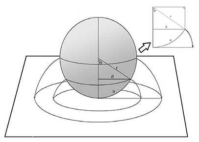
To develop a novel, simplified method for correcting the ischemic index of nonperfused areas in diabetic retinopathy (DR).
METHODS
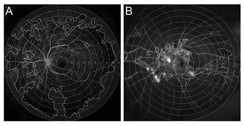
We performed a retrospective review of 103 eyes with naive DR that underwent ultra-widefield angiography (UWFA) over a year. UWFAs were graded according to the quantity of retinal non-perfusion, and uncorrected ischemic index (UII) and corrected ischemic index (CII) were calculated using a simplified, novel method.
RESULTS
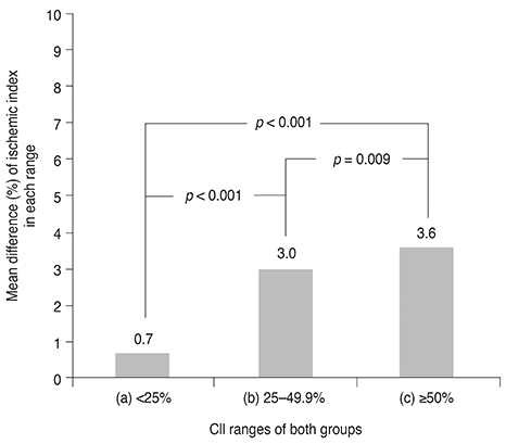
The average differences between UII and CII in the non-proliferative DR group and the proliferative DR group were 0.7 +/- 0.9% in the <25% CII group, 3.0 +/- 0.9% in the 25% to 49.9% CII group, and 3.6 +/- 0.6% in the >50% CII group, respectively. A CII >25% was critical for determining DR progression (p < 0.001).
CONCLUSIONS
Distortion created by UWFA needs to be corrected because the difference between UII and CII in DR increases with the ischemic index.
MeSH Terms
Figure
Reference
-
1. Wessel MM, Aaker GD, Parlitsis G, et al. Ultra-wide-field angiography improves the detection and classification of diabetic retinopathy. Retina. 2012; 32:785–791.2. Tsui I, Kaines A, Havunjian MA, et al. Ischemic index and neovascularization in central retinal vein occlusion. Retina. 2011; 31:105–110.3. Patel RD, Messner LV, Teitelbaum B, et al. Characterization of ischemic index using ultra-widefield fluorescein angiography in patients with focal and diffuse recalcitrant diabetic macular edema. Am J Ophthalmol. 2013; 155:1038–1044.4. Prasad PS, Oliver SC, Coffee RE, et al. Ultra wide-field angiographic characteristics of branch retinal and hemicentral retinal vein occlusion. Ophthalmology. 2010; 117:780–784.5. Silva PS, Cavallerano JD, Sun JK, et al. Nonmydriatic ultrawide field retinal imaging compared with dilated standard 7-field 35-mm photography and retinal specialist examination for evaluation of diabetic retinopathy. Am J Ophthalmol. 2012; 154:549–559.6. Oliver SC, Schwartz SD. Peripheral vessel leakage (PVL): a new angiographic finding in diabetic retinopathy identified with ultra wide-field fluorescein angiography. Semin Ophthalmol. 2010; 25:27–33.7. Spaide RF. Peripheral areas of nonperfusion in treated central retinal vein occlusion as imaged by wide-field fluorescein angiography. Retina. 2011; 31:829–837.8. Wessel MM, Nair N, Aaker GD, et al. Peripheral retinal ischaemia, as evaluated by ultra-widefield fluorescein angiography, is associated with diabetic macular oedema. Br J Ophthalmol. 2012; 96:694–698.9. Oishi A, Hidaka J, Yoshimura N. Quantification of the image obtained with a wide-field scanning ophthalmoscope. Invest Ophthalmol Vis Sci. 2014; 55:2424–2431.
- Full Text Links
- Actions
-
Cited
- CITED
-
- Close
- Share
- Similar articles
-
- Anterior Diabetic Retinopathy Studied by Ultra-widefield Angiography
- Correlations between Macular Microvascular Alterations and Peripheral Ischemia in Patients with Branch Retinal Vein Occlusion
- The Effect of Fluorescein Angiography on Renal Function in Patients with Diabetic Retinopathy
- Ultrawide-field Fluorescein Angiography for Evaluation of Diabetic Retinopathy
- Clinical Analysis of Diabetic Retinopathy According to the Type of Diabetes Mellitus


