J Korean Foot Ankle Soc.
2016 Dec;20(4):192-195. 10.14193/jkfas.2016.20.4.192.
Pigmented Villonodular Synovitis of the Ankle and Subtalar Joint Treated by Surgical Excision and Ligament Reconstructions: A Case Report
- Affiliations
-
- 1Department of Orthopedic Surgery, Kangdong Sacred Heart Hospital, Hallym University College of Medicine, Seoul, Korea. kiga9@hanmail.net
- KMID: 2362769
- DOI: http://doi.org/10.14193/jkfas.2016.20.4.192
Abstract
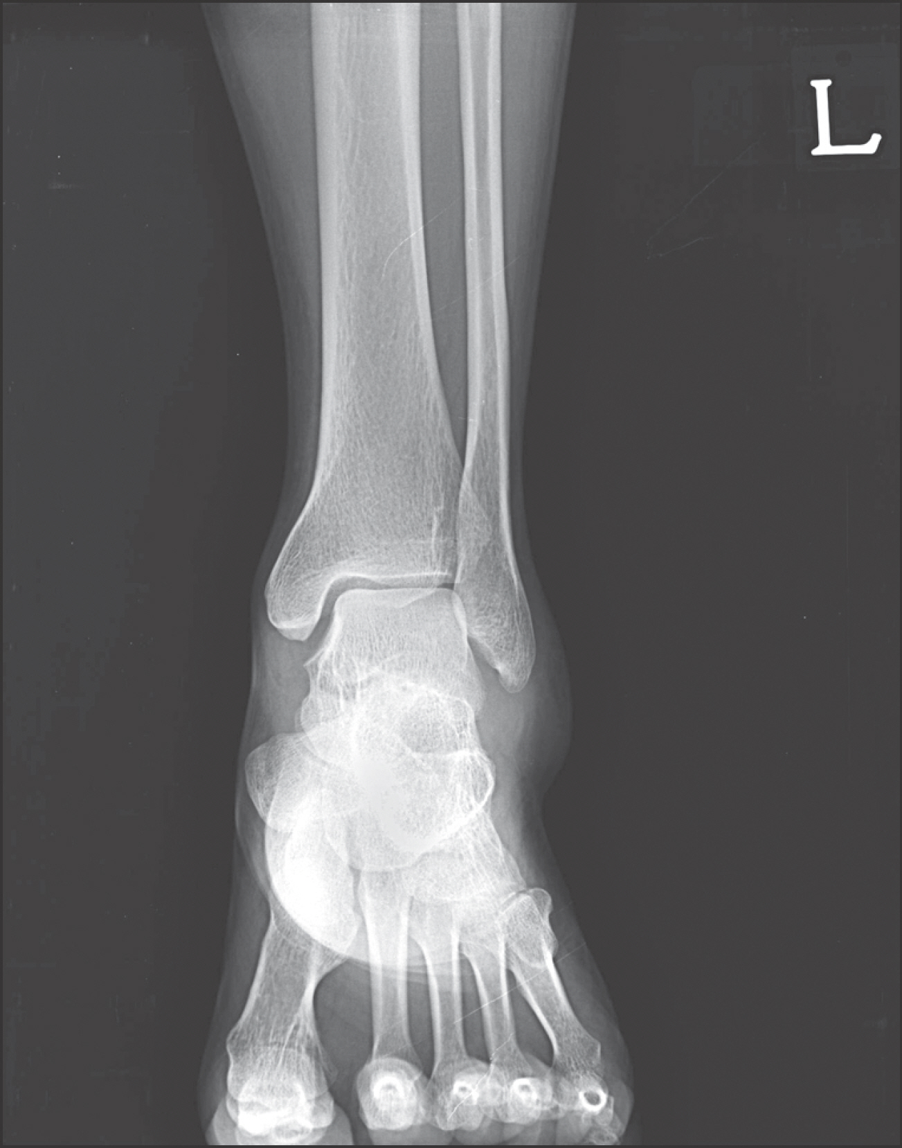
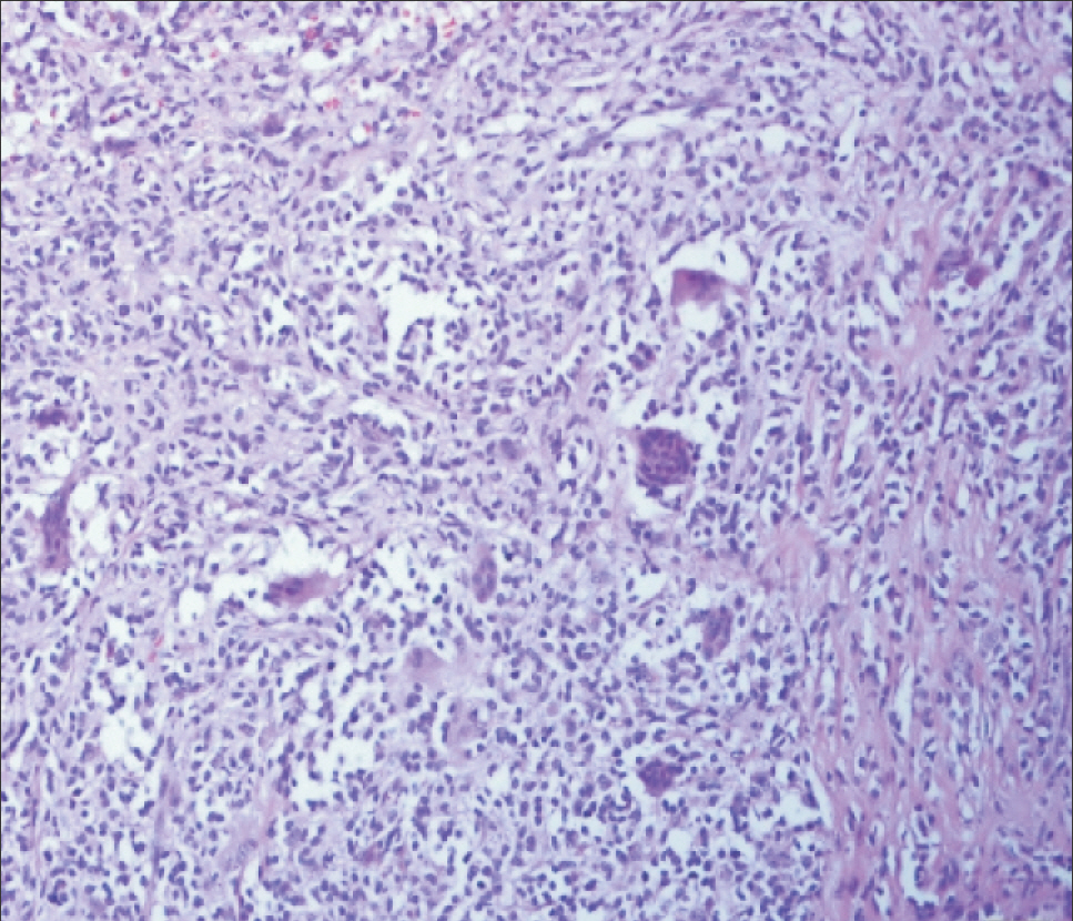
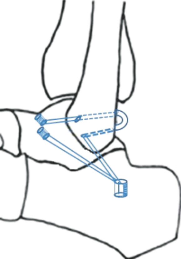
- Diffuse pigmented villonodular synovitis (PVNS) involving ankle joint needs complete mass excision and total synovectomy to reduce recurrence rate, while surrounding ligaments can be easily damaged. So the concurrent ligament reconstruction should be considered for post-excisional instability in subtalar joint as well as lateral ankle joint. We describe our experience in the management of a diffuse type PVNS, invades lateral talocrural joint extended to subtalar joint and introduce a new technique of all-in-one reconstruction for anterior talofibular,calcaneofibular and cervical ligament. Our new reconstruction technique applying modified Chrisman and Snook technique is useful in stabilization for deficiencies of the ligament complexafter PVNS excisionat lateral ankle and subtalar joint.
Keyword
MeSH Terms
Figure
Cited by 1 articles
-
Natural History of Osteochondral Lesion of the Talus
Min Gyu Kyung, Dong-Oh Lee, Dong Yeon Lee
J Korean Foot Ankle Soc. 2020;24(2):37-41. doi: 10.14193/jkfas.2020.24.2.37.
Reference
-
1.Stevenson JD., Jaiswal A., Gregory JJ., Mangham DC., Cribb G., Cool P. Diffuse pigmented villonodular synovitis (diffuse-type giant cell tumour) of the foot and ankle. Bone Joint J. 2013. 95:384–90.
Article2.Frassica FJ., Bhimani MA., McCarthy EF., Wenz J. Pigmented villonodular synovitis of the hip and knee. Am Fam Physician. 1999. 60:1404–10.3.Hertel J. Functional anatomy, pathomechanics, and pathophysiology of lateral ankle instability. J Athl Train. 2002. 37:364–75.4.Schnirring-Judge M., Lin B. Pigmented villonodular synovitis of the ankle-radiation therapy as a primary treatment to reduce recurrence: a case report with 8-year follow-up. J Foot Ankle Surg. 2011. 50:108–16.
Article5.Ward WG Sr., Boles CA., Ball JD., Cline MT. Diffuse pigmented villonodular synovitis: preliminary results with intralesional resection and p32 synoviorthesis. Clin Orthop Relat Res. 2007. 454:186–91.6.Elmslie RC. Recurrent subluxation of the ankle-joint. Ann Surg. 1934. 100:364–7.7.Chrisman OD., Snook GA. Reconstruction of lateral ligament tears of the ankle. An experimental study and clinical evaluation of seven patients treated by a new modification of the Elmslie procedure. J Bone Joint Surg Am. 1969. 51:904–12.8.Karlsson J., Eriksson BI., Renström P. Subtalar instability of the foot. A review and results after surgical treatment. Scand J Med Sci Sports. 1998. 8:191–7.
Article9.Aynardi M., Pedowitz DI., Raikin SM. Subtalar instability. Foot Ankle Clin. 2015. 20:243–52.
Article10.Gould N., Seligson D., Gassman J. Early and late repair of lateral ligament of the ankle. Foot Ankle. 1980. 1:84–9.
Article
- Full Text Links
- Actions
-
Cited
- CITED
-
- Close
- Share
- Similar articles
-
- Total Ankle Replacement in Pigmented Villonodular Synovitis of Ankle Joint (A Case Report)
- Nodular Pigmented Villonodular Synovitis of the Right Shoulder Joint: One Case Report
- Simultaneous Pigmented Villonodular Synovitis and Synovial Chondromatosis in the Ankle Joint
- Open Synovectomy in Diffuse Pigmented Villonodular Synovitis of Ankle Joint: A Case Report
- A Case of Pigmented Villonodular Synovitis


