Change of Reliability for Distal Metatarsal Articular Angle Measurement before and after Proximal Chevron Osteotomy
- Affiliations
-
- 1Department of Orthopedic Surgery, Yeungnam University College of Medicine, Daegu, Korea. chpark77@naver.com
- KMID: 2362761
- DOI: http://doi.org/10.14193/jkfas.2016.20.4.145
Abstract
- PURPOSE
To evaluate the reliability of preoperative and postoperative distal metatarsal articular angle (DMAA) measurements and to determine whether such reliability is different in accordance with the foot and ankle fellowship and the number of years in practice.
MATERIALS AND METHODS
Between July 2012 and June 2014, a total of 20 patients (24 feet) were treated with proximal chevron osteotomy and distal soft tissue procedure for symptomatic hallux valgus deformity. DMAA were measured twice with an interval of two weeks between the preoperative and postoperative dorsoplantar radiographs by four observers; two of whom were foot and ankle surgeons (A and B), one knee surgeon, and one senior resident. The intraobserver reproducibility and interobserver reliability were assessed by intraclass correlation coefficients. Moreover, the limit of agreement between the preoperative and postoperative DMAA measurements were assessed using a Bland-Altman plot.
RESULTS
The intraobserver reproducibility of the foot and ankle surgeon A, knee surgeon, and senior resident improved from 0.796, 0.575, and 0.586 preoperatively to 0.968, 0.864, and 0.864 postoperatively, respectively. The interobserver reliability of foot and ankle surgeon A-B, foot and ankle surgeon A-knee surgeon, and foot and ankle surgeon A-senior resident improved from 0.874, 0.688, and 0.677 preoperatively to 0.971, 0.917, and 0.838 postoperatively, respectively.
CONCLUSION
The intra- and interobserver reliabilities for DMAA measurement improved after proximal chevron osteotomy. Therefore, the necessity of additional procedures to correct the increased DMAA should be reevaluated after proximal chevron osteotomy in the hallux valgus with an increased DMAA.
MeSH Terms
Figure
-
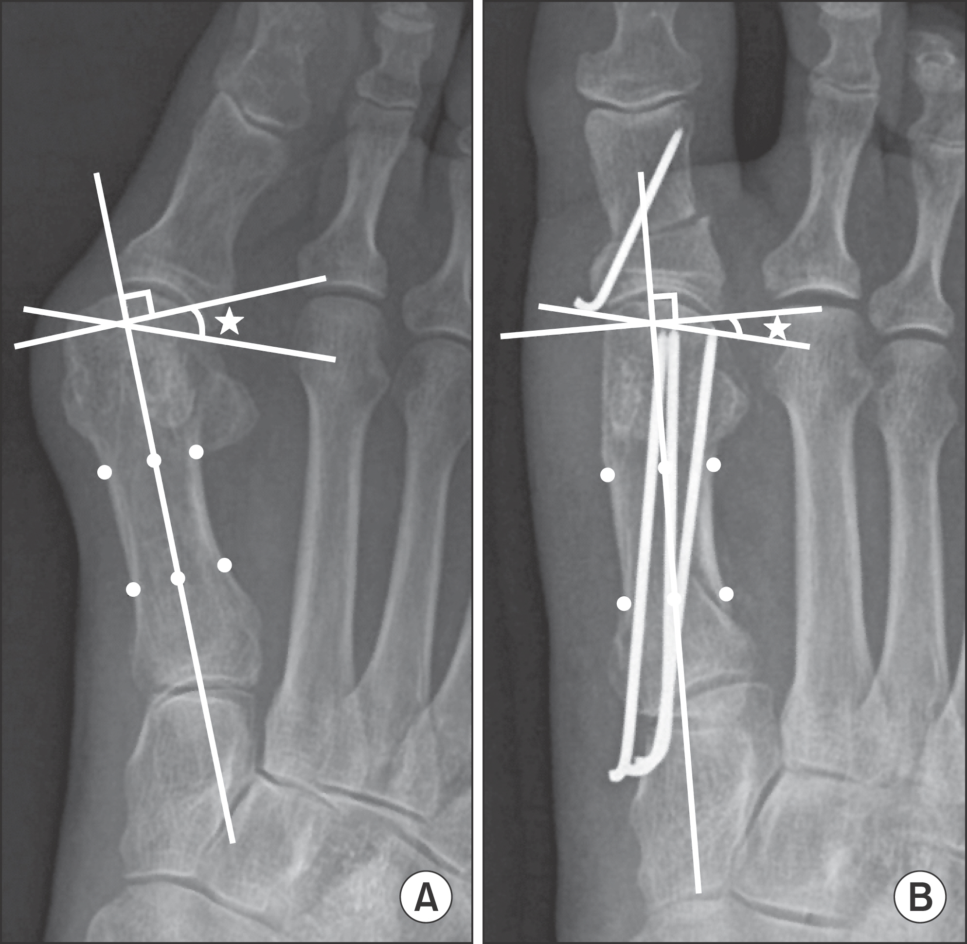
Figure 1. Distal metatarsal articular angle is measured on preoperative (A) and postoperative (B) radiographs. Distal metatarsal articular angle (asterisks) defined as the angle between a perpendicular line to the longitudinal axis of the first metatarsal and a line delineating the orientation of the metatarsal head articular surface.

Figure 2. Graphs show Bland-Altman plot of the difference of distal metatarsal articular angle measurement of foot and ankle surgeon A on preoperative (A) and postoperative (B) radiographs. DMAA: distal metatarsal articular angle.
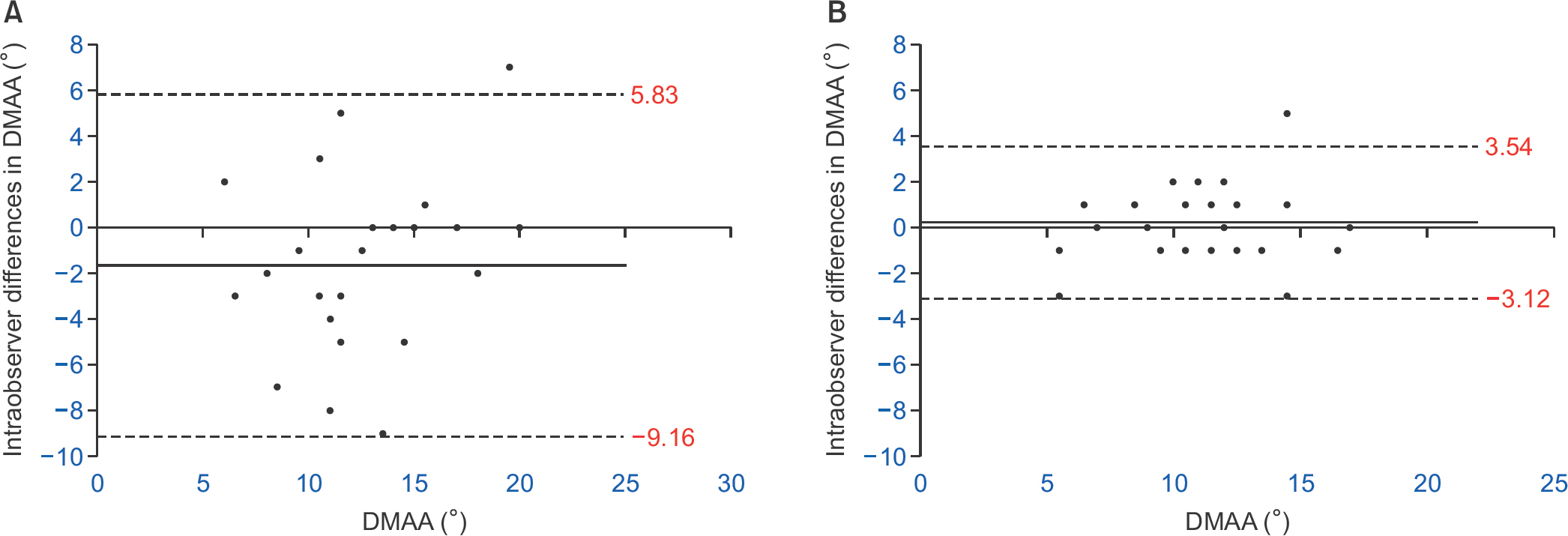
Figure 3. Graphs show Bland-Altman plot of the difference of distal metatarsal articular angle measurement of knee surgeon on preoperative (A) and postoperative (B) radiographs. DMAA: distal metatarsal articular angle.

Figure 4. Graphs show Bland-Altman plot of the difference of distal metatarsal articular angle measurement of senior resident on preoperative (A) and postoperative (B) radiographs. DMAA: distal metatarsal articular angle.
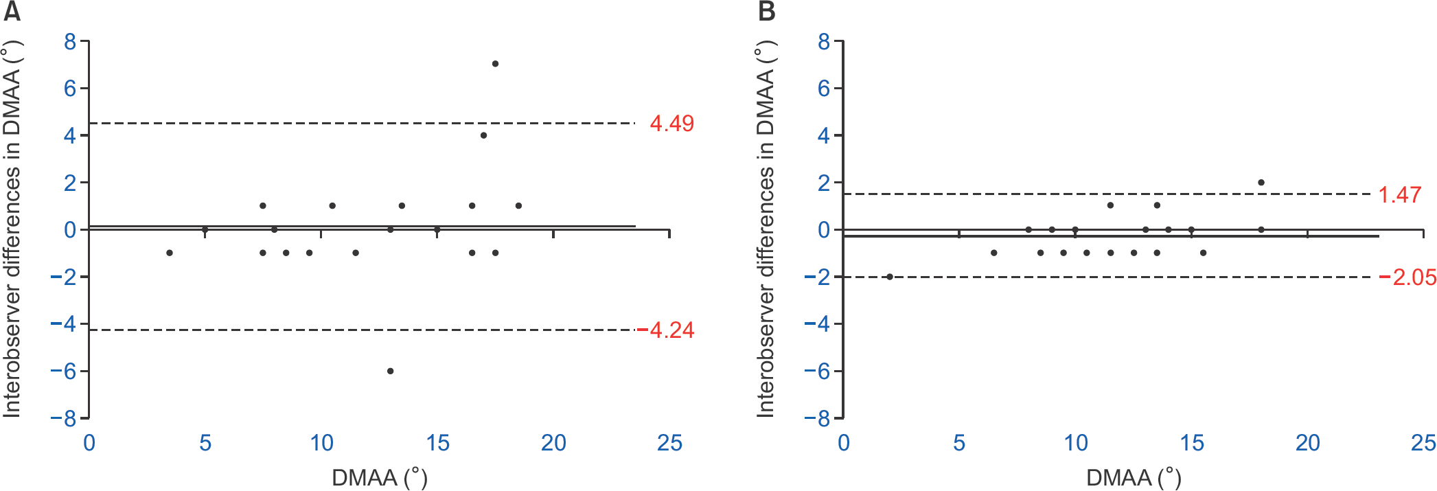
Figure 5. Graphs show Bland-Altman plot of the difference of distal metatarsal articular angle measurement between foot and ankle surgeon A and B on preoperative (A) and postoperative (B) radiographs. DMAA: distal metatarsal articular angle.
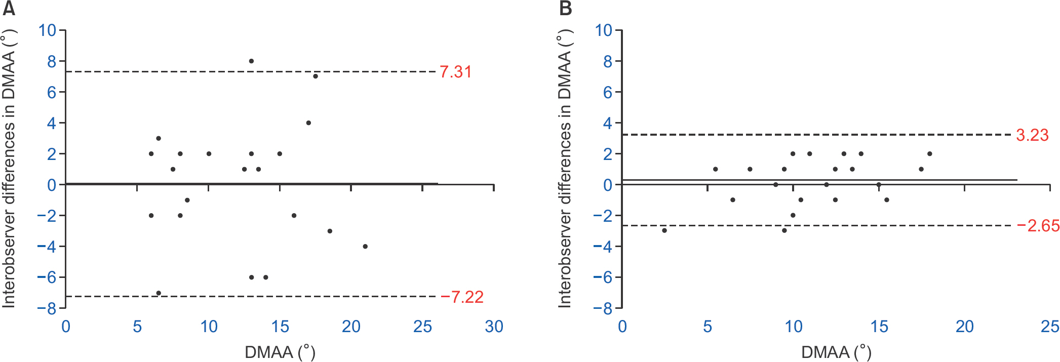
Figure 6. Graphs show Bland-Altman plot of the difference of distal metatarsal articular angle measurement between foot and ankle surgeon A and knee surgeon on preoperative (A) and postoperative (B) radiographs. DMAA: distal metatarsal articular angle.

Figure 7. Graphs show Bland-Altman plot of the difference of distal metatarsal articular angle measurement between foot and ankle surgeon A and senior resident on preoperative (A) and postoperative (B) radiographs. DMAA: distal metatarsal articular angle.
Cited by 1 articles
-
Radiographic Risk Factors of Recurrent Hallux Valgus Deformity after Modified Scarf and Akin Osteotomy
Jae Wan Suh, Sung Hyun Kim, Hyun-Woo Park
J Korean Foot Ankle Soc. 2019;23(4):159-165. doi: 10.14193/jkfas.2019.23.4.159.
Reference
-
1.Bonnel F., Canovas F., Poirée G., Dusserre F., Vergnes C. Evaluation of the scarf osteotomy in hallux valgus related to distal metatarsal articular angle: a prospective study of 79 operated cases. Rev Chir Orthop Reparatrice Appar Mot. 1999. 85:381–6.2.Park CH., Cho JH., Moon JJ., Lee WC. Can double osteotomy be a solution for adult hallux valgus deformity with an increased distal metatarsal articular angle? J Foot Ankle Surg. 2016. 55:188–92.
Article3.Chi TD., Davitt J., Younger A., Holt S., Sangeorzan BJ. Intra- and inter-observer reliability of the distal metatarsal articular angle in adult hallux valgus. Foot Ankle Int. 2002. 23:722–6.
Article4.Coughlin MJ. Roger A1 Mann Award1 Juvenile hallux valgus: etiology and treatment. Foot Ankle Int. 1995. 16:682–97.5.Okuda R., Kinoshita M., Yasuda T., Jotoku T., Kitano N., Shima H. Postoperative incomplete reduction of the sesamoids as a risk factor for recurrence of hallux valgus. J Bone Joint Surg Am. 2009. 91:1637–45.
Article6.Lee KM., Ahn S., Chung CY., Sung KH., Park MS. Reliability and relationship of radiographic measurements in hallux valgus. Clin Orthop Relat Res. 2012. 470:2613–21.
Article7.Bland JM., Altman DG. Statistical methods for assessing agreement between two methods of clinical measurement. Lancet. 1986. 1:307–10.
Article8.Shrout PE., Fleiss JL. Intraclass correlations: uses in assessing rater reliability. Psychol Bull. 1979. 86:420–8.
Article9.Eustace S., O’Byrne J., Stack J., Stephens MM. Radiographic features that enable assessment of first metatarsal rotation: the role of pronation in hallux valgus. Skeletal Radiol. 1993. 22:153–6.
Article10.Coughlin MJ., Freund E. The reliability of angular measurements in hallux valgus deformities. Foot Ankle Int. 2001. 22:369–79.
Article11.Chou LB., Mann RA., Casillas MM. Biplanar chevron osteotomy. Foot Ankle Int. 1998. 19:579–84.
Article12.Corte-Real NM., Moreira RM. Modified biplanar chevron oste-otomy. Foot Ankle Int. 2009. 30:1149–53.
Article13.DeOrio J. Technique tip: dorsal wedge resection (uniplanar) in the chevron osteotomy for high distal metatarsal articular angle bunions. Foot Ankle Int. 2007. 28:642–4.
Article14.Aronson J., Nguyen LL., Aronson EA. Early results of the modified peterson bunion procedure for adolescent hallux valgus. J Pediatr Orthop. 2001. 21:65–9.
Article15.Coughlin MJ., Carlson RE. Treatment of hallux valgus with an increased distal metatarsal articular angle: evaluation of double and triple first ray osteotomies. Foot Ankle Int. 1999. 20:762–70.
Article16.Johnson AE., Georgopoulos G., Erickson MA., Eilert R. Treatment of adolescent hallux valgus with the first metatarsal double osteotomy: the denver experience. J Pediatr Orthop. 2004. 24:358–62.17.Yasuda T., Okuda R., Jotoku T., Shima H., Hida T., Neo M. Proximal supination osteotomy of the first metatarsal for hallux valgus. Foot Ankle Int. 2015. 36:696–704.
Article18.Scott G., Wilson DW., Bentley G. Roentgenographic assessment in hallux valgus. Clin Orthop Relat Res. 1991. 267:143–7.
Article19.Shereff MJ., DiGiovanni L., Bejjani FJ., Hersh A., Kummer FJ. A comparison of nonweight-bearing and weight-bearing radiographs of the foot. Foot Ankle. 1990. 10:306–11.
Article20.Tanaka Y., Takakura Y., Takaoka T., Akiyama K., Fujii T., Tamai S. Radiographic analysis of hallux valgus in women on weightbearing and nonweightbearing. Clin Orthop Relat Res. 1997. 336:186–94.
Article21.Robinson AH., Cullen NP., Chhaya NC., Sri-Ram K., Lynch A. Variation of the distal metatarsal articular angle with axial rotation and inclination of the first metatarsal. Foot Ankle Int. 2006. 27:1036–40.
Article22.Vittetoe DA., Saltzman CL., Krieg JC., Brown TD. Validity and reliability of the first distal metatarsal articular angle. Foot Ankle Int. 1994. 15:541–7.
Article
- Full Text Links
-
- Actions
-
Cited
- CITED
-
- Close
- Share
-
- Similar articles
-
- Comparison of Proximal Metatarsal Osteotomy andDistal Chevron Osteotomy for Correction of Hallux Valgus
- Treatment of Hallux Valgus with Distal Chevron Metatarsal Osteotomy
- Chevron Osteotomy as the Treatment of Moderate to Severe Hallux Valgus Deformity
- Comparison of the Results between Distal Chevron Osteotomy and Proximal Metatarsal Osteotomy for the Treatment of Moderate Hallux Valgus
- Radiological Comparison between 60 degrees Distal Chevron Osteotomy and 40 degrees Distal Chevron Osteotomy in Hallux Valgus
