J Korean Soc Radiol.
2016 Dec;75(6):487-494. 10.3348/jksr.2016.75.6.487.
Detection of Parenchymal Abnormalities in Experimentally Induced Acute Pyelonephritis in Rabbits Using Contrast-Enhanced Ultrasonography, CT, and MRI
- Affiliations
-
- 1Department of Radiology, Samsung Medical Center, Sungkyunkwan University School of Medicine, Seoul, Korea. ryuja@hanyang.ac.kr
- 2Department of Radiology, Guri Hospital, Hanyang University College of Medicine, Guri, Korea.
- 3Department of Radiology, Mayo Clinic College of Medicine, Rochester, MN, USA.
- 4Electronic Radiology Laboratory, Department of Radiology, Mallinckrodt Institute of Radiology, Washington University School of Medicine, St. Louis, MO, USA.
- 5Department of Pathology, Samsung Medical Center, Sungkyunkwan University School of Medicine, Seoul, Korea.
- 6Department of Pathology, Sacred Heart Hospital, Hallym University College of Medicine, Anyang, Korea.
- 7Laboratory Animal Research Center, Samsung Biomedical Research Institute, Sungkyunkwan University School of Medicine, Seoul, Korea.
- KMID: 2360437
- DOI: http://doi.org/10.3348/jksr.2016.75.6.487
Abstract
- PURPOSE
We evaluated the efficacy of contrast-enhanced ultrasonography (CEUS) in detecting acute pyelonephritis (APN) using the rabbit kidney model and compared it with CT and MRI.
MATERIALS AND METHODS
This study was approved by the Institutional Review Board. In a total of 20 New Zealand White rabbits, APN was induced experimentally. CEUS, CT, and MRI were performed on the first, third, and seventh postoperative days. After imaging studies, the subjects were sacrificed and the pathological diagnosis of APN was confirmed in each animal by a pathologist.
RESULTS
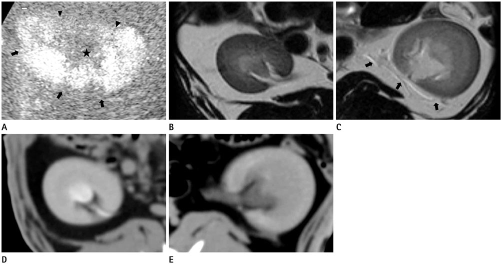
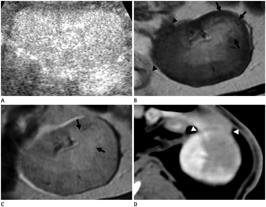
Imaging studies were obtained in eight animals, including eight CEUS, four computed tomography (CT), and four magnetic resonance imaging (MRI) images. CEUS depicted diffuse renal enlargement (7), diffuse heterogeneous parenchymal enhancement (6), and focal areas of decreased parenchymal enhancement (6). These findings were well correlated with the CT and MRI findings in five cases in which these studies were available. CT and MRI showed diffuse renal enlargement, diffuse heterogeneous parenchymal enhancement, focal areas of decreased parenchymal enhancement, focal contour bulging, and the finding of perinephric spread of infection.
CONCLUSION
In a rabbit model, CEUS could depict the parenchymal lesions of APN similar to CT or MRI; however, it was limited in depicting the perinephric extension of inflammation.
MeSH Terms
Figure
Reference
-
1. Dacher JN, Pfister C, Monroc M, Eurin D, LeDosseur P. Power Doppler sonographic pattern of acute pyelonephritis in children: comparison with CT. AJR Am J Roentgenol. 1996; 166:1451–1455.2. Stunell H, Buckley O, Feeney J, Geoghegan T, Browne RF, Torreggiani WC. Imaging of acute pyelonephritis in the adult. Eur Radiol. 2007; 17:1820–1828.3. Craig WD, Wagner BJ, Travis MD. Pyelonephritis: radiologic-pathologic review. Radiographics. 2008; 28:255–277. quiz 327-328.4. Kim B, Lim HK, Choi MH, Woo JY, Ryu J, Kim S, et al. Detection of parenchymal abnormalities in acute pyelonephritis by pulse inversion harmonic imaging with or without microbubble ultrasonographic contrast agent: correlation with computed tomography. J Ultrasound Med. 2001; 20:5–14.5. Fontanilla T, Minaya J, Cortés C, Hernando CG, Arangüena RP, Arriaga J, et al. Acute complicated pyelonephritis: contrast-enhanced ultrasound. Abdom Imaging. 2012; 37:639–646.6. Taylor GA, Barnewolt CE, Claudon M, Dunning PS. Depiction of renal perfusion defects with contrast-enhanced harmonic sonography in a porcine model. AJR Am J Roentgenol. 1999; 173:757–760.7. Claudon M, Barnewolt CE, Taylor GA, Dunning PS, Gobet R, Badawy AB. Renal blood flow in pigs: changes depicted with contrast-enhanced harmonic US imaging during acute urinary obstruction. Radiology. 1999; 212:725–731.8. Pennington DJ, Lonergan GJ, Flack CE, Waguespack RL, Jackson CB. Experimental pyelonephritis in piglets: diagnosis with MR imaging. Radiology. 1996; 201:199–205.9. Runge VM, Timoney JF, Williams NM. Magnetic resonance imaging of experimental pyelonephritis in rabbits. Invest Radiol. 1997; 32:696–704.10. Giamarellos-Bourboulis E, Adamis T, Sabracos L, Raftogiannis M, Baziaka F, Tsaganos T, et al. Clarithromycin: immunomodulatory therapy of experimental sepsis and acute pyelonephritis by Escherichia coli. Scand J Infect Dis. 2005; 37:48–54.
- Full Text Links
- Actions
-
Cited
- CITED
-
- Close
- Share
- Similar articles
-
- Local ablation therapy with contrast-enhanced ultrasonography for hepatocellular carcinoma: a practical review
- Unraveling distinctions between contrast-enhanced ultrasound and CT/MRI for liver mass diagnosis
- Acute pyelonephritis focusing on perfusion defects on contrast enhanced computerized tomography(CT) scans and its clinical outcome
- Diagnostic Performance of Gray-scale Sonographic Findings for the Detection of Acute Pyelonephritis
- CT Findings of Acute Pyelonephritis in Children:Correlation with Clinical Manifestations


