Clin Orthop Surg.
2015 Dec;7(4):483-489. 10.4055/cios.2015.7.4.483.
Intraoperative Three-Dimensional Imaging in Calcaneal Fracture Treatment
- Affiliations
-
- 1Department of Orthopaedic Surgery, Inje University Busan Paik Hospital, Inje University College of Medicine, Busan, Korea.
- 2Department of Orthopedics, District Hospital, Korea Army Training Center, Nonsan, Korea. bluewhisle@gmail.com
- KMID: 2360263
- DOI: http://doi.org/10.4055/cios.2015.7.4.483
Abstract
- BACKGROUND
To compare the effectiveness of intraoperative three-dimensional (3D) image and conventional two-dimensional (2D) fluoroscopic images, which are used in the treatment of acute calcaneal fractures.
METHODS
We retrospectively analyzed 40 patients who suffered calcaneal fracture and underwent surgery at Inje University Busan Paik Hospital. The patients were divided into two groups. Only 2D fluoroscopy was used to evaluate 20 patients of group 1. On the other hand, 3D fluoroscopy was performed on the remaining 20 patients of group 2; 3D fluoroscopy was performed on these patients after they were extensively evaluated by 2D fluoroscopy during surgery. We reviewed the radiographic and clinical outcomes of these patients, whose average follow-up period was 42.6 months.
RESULTS
In group 2, 3D fluoroscopy detected four cases (20%) of articular incongruence and screw misplacement. All these complicated cases were corrected during surgery. At the final follow-up session, the mean American Orthopedic Foot and Ankle Society (AOFAS) hind foot score was 78.3 (range, 65 to 95) in group 1 and 82.3 (range, 68 to 95) in group 2.
CONCLUSIONS
Intraoperative 3D imaging of calcaneal fractures is considered to be useful in evaluating the congruence of joints and the placement of implants.
Keyword
MeSH Terms
Figure
-
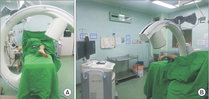
Fig. 1 Three-dimensional fluoroscopy. (A) Draping was performed using a sterile cover for fluoroscopes. (B) The device performs a 190-degree orbital rotation within two minutes.
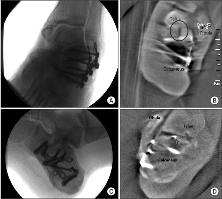
Fig. 2 Cases in which the screw was modified. (A, C) After evaluation with two-dimensional fluoroscopy, reduction and implant placement were checked by the surgeon. (B, D) Three-dimensional fluoroscopy showed a step-off and gap of the fracture site and wrong position of the screw (arrow), which were corrected.
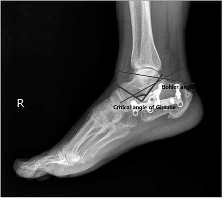
Fig. 3 Lateral radiograph of the calcaneus shows the measurements of the critical angle of Bohler's and Gissane's angle.
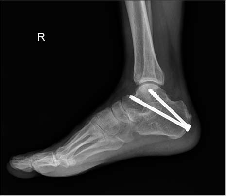
Fig. 4 A case with complication. After treating the deep infection, we performed subtalar fusion.
Reference
-
1. Hufner T, Stubig T, Gosling T, Kendoff D, Geerling J, Krettek C. Cost-benefit analysis of intraoperative 3D imaging. Unfallchirurg. 2007; 110(1):14–21.2. Richter M, Geerling J, Zech S, Goesling T, Krettek C. Intraoperative three-dimensional imaging with a motorized mobile C-arm (SIREMOBIL ISO-C-3D) in foot and ankle trauma care: a preliminary report. J Orthop Trauma. 2005; 19(4):259–266.
Article3. Rubberdt A, Feil R, Stengel D, et al. The clinical use of the ISO-C(3D) imaging system in calcaneus fracture surgery. Unfallchirurg. 2006; 109(2):112–118.4. Gavlik JM, Rammelt S, Zwipp H. The use of subtalar arthroscopy in open reduction and internal fixation of intra-articular calcaneal fractures. Injury. 2002; 33(1):63–71.
Article5. Sanders R, Gregory P. Operative treatment of intra-articular fractures of the calcaneus. Orthop Clin North Am. 1995; 26(2):203–214.
Article6. Benirschke SK, Sangeorzan BJ. Extensive intraarticular fractures of the foot: surgical management of calcaneal fractures. Clin Orthop Relat Res. 1993; (292):128–134.7. Bezes H, Massart P, Delvaux D, Fourquet JP, Tazi F. The operative treatment of intraarticular calcaneal fractures: indications, technique, and results in 257 cases. Clin Orthop Relat Res. 1993; (290):55–59.8. Carr JB. Surgical treatment of the intra-articular calcaneus fracture. Orthop Clin North Am. 1994; 25(4):665–675.
Article9. Leung KS, Yuen KM, Chan WS. Operative treatment of displaced intra-articular fractures of the calcaneum: medium-term results. J Bone Joint Surg Br. 1993; 75(2):196–201.
Article10. Paley D, Hall H. Intra-articular fractures of the calcaneus: a critical analysis of results and prognostic factors. J Bone Joint Surg Am. 1993; 75(3):342–354.
Article11. Zwipp H, Tscherne H, Thermann H, Weber T. Osteosynthesis of displaced intraarticular fractures of the calcaneus: results in 123 cases. Clin Orthop Relat Res. 1993; (290):76–86.12. Sanders R, Fortin P, DiPasquale T, Walling A. Operative treatment in 120 displaced intraarticular calcaneal fractures: results using a prognostic computed tomography scan classification. Clin Orthop Relat Res. 1993; (290):87–95.13. Sanders R, Fortin P, DiPasquale T, Walling A, Helfet D, Ross E. The results of operative treatment of displaced intra-articular calcaneal fractures using a CT scan classification. In : Tscherne H, Schatzker J, editors. Major fractures of the pilon, the talus, and the calcaneus: current concepts of treatment. Berlin: Springer;1993. p. 175–189.14. Trepman E, Lutter LD, Brodsky JW. Highlights of the Fourteenth Annual Summer Meeting of the American Orthopaedic Foot and Ankle Society, Boston, Massachusetts, July 24-26, 1998. Foot Ankle Int. 1999; 20(1):58–67.
Article15. Jordan C, Mirzabeigi E, Williams S. Determining the angle of screw placement for internal fixation of calcaneal fractures. J Orthop Trauma. 1999; 13(1):47–50.
Article16. Euler E, Wirth S, Linsenmaier U, Mutschler W, Pfeifer KJ, Hebecker A. Comparative study of the quality of C-arm based 3D imaging of the talus. Unfallchirurg. 2001; 104(9):839–846.17. Kotsianos D, Wirth S, Fischer T, et al. 3D imaging with an isocentric mobile C-arm comparison of image quality with spiral CT. Eur Radiol. 2004; 14(9):1590–1595.18. Rock C, Kotsianos D, Linsenmaier U, et al. Studies on image quality, high contrast resolution and dose for the axial skeleton and limbs with a new, dedicated CT system (ISO-C-3 D). Rofo. 2002; 174(2):170–176.
Article19. Rock C, Linsenmaier U, Brandl R, et al. Introduction of a new mobile C-arm/CT combination equipment (ISO-C-3D): initial results of 3-D sectional imaging. Unfallchirurg. 2001; 104(9):827–833.20. Stockle U, Krettek C, Pohlemann T, Messmer P. Clinical applications: pelvis. Injury. 2004; 35:Suppl 1. S-A46–S-A56.21. Wich M, Spranger N, Ekkernkamp A. Intraoperative imaging with the ISO C(3D). Chirurg. 2004; 75(10):982–987.
- Full Text Links
-
- Actions
-
Cited
- CITED
-
- Close
- Share
-
- Similar articles
-
- Atraumatic Avulsion Fracture of Calcaneal Tuberosity in a Patient with Peripheral Neuropathy: A Case Report
- Avulsion Fracture of Calcaneal Tubercle Treated with Cannulated Cancellous Screws and Wire: Surgical Technique
- Operative Treatment of Calcaneal Fracture
- The Fate of Fracture Fragment in Diabetic Calcaneal Insufficiency Avulsion Fracture
- The Effect of External Distractor on Recovery of B hler angle in Displaced Intraarticular Calcaneal Fractures
