Uterine Intravenous Leiomyomatosis with Intracardiac Extension and Pulmonary Benign Metastases on FDG PET/CT: A Case Report
- Affiliations
-
- 1PET/CT Center, Gansu Provincial Hospital, Lanzhou, Gansu 730000, China. gssypetct@163.com
- KMID: 2360216
- DOI: http://doi.org/10.3348/kjr.2016.17.2.289
Abstract
- A 48-year-old woman presented with a 50-day history of irregular vaginal bleeding and lower abdominal pain. Ultrasound indicated an extremely large occupying lesion in the pelvic cavity that was highly suggestive of malignancy. Fluorodeoxyglucose (FDG) positron emission tomography/computed tomography (PET/CT) was performed to further assess the nature of pelvic abnormality. PET/CT images demonstrated a diffusely lobulated mass ranging from cervix up to the inferior pole of kidneys with mild FDG uptake. Simultaneously, multiple nodules in bilateral lungs and a hypodense lesion in the right ventricle were shown without FDG-avidity. Based on the imaging results, the presumptive diagnosis was uterine intravenous leiomyomatosis with intracardiac extension and pulmonary benign metastases, which was subsequently confirmed by MRI and the lesion biopsy.
MeSH Terms
-
Female
Fluorodeoxyglucose F18/chemistry
Humans
Leiomyoma/pathology/radiography
Leiomyomatosis/pathology/*radiography
Lung Neoplasms/radiography/*secondary
Magnetic Resonance Imaging
Middle Aged
Positron-Emission Tomography
Tomography, X-Ray Computed
Uterine Neoplasms/pathology/radiography
Vena Cava, Inferior/pathology
Fluorodeoxyglucose F18
Figure
-
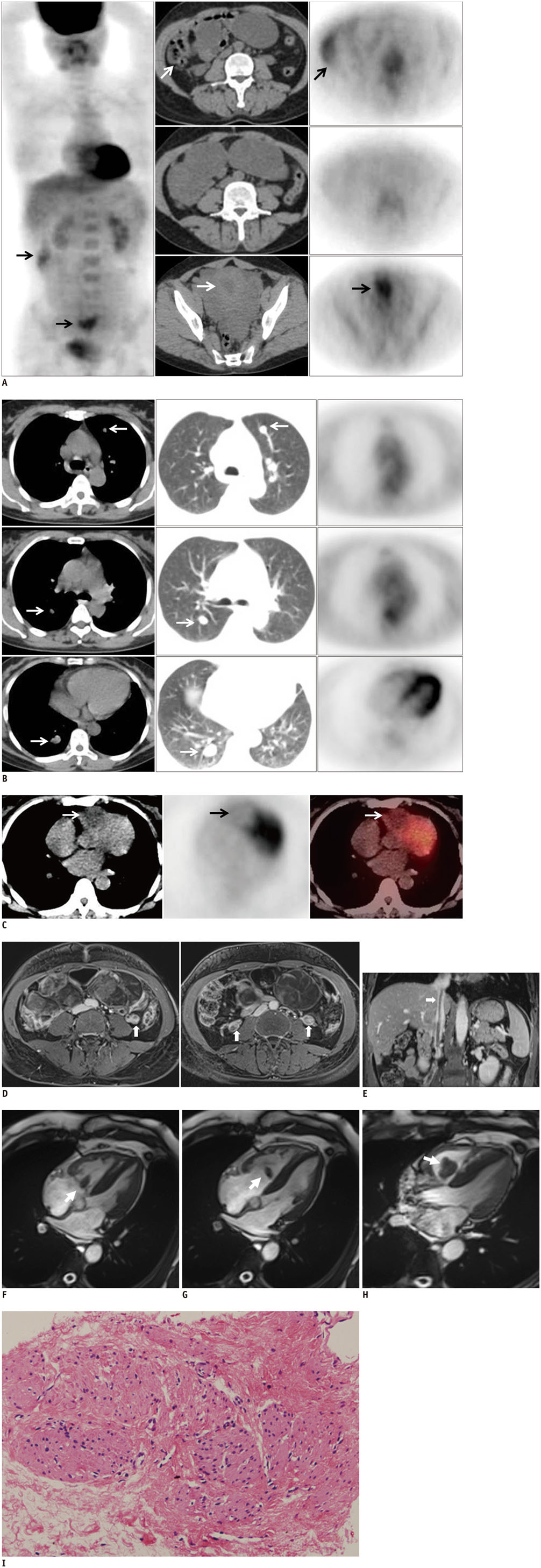
Fig. 1 48-year-old woman with uterine intravenous leiomyomatosis accompanied by intracardiac extension and pulmonary benign metastases. A. MIP image of FDG PET, and transverse FDG PET/CT images of abdomen showed physiological FDG uptake in colon and endometrium (arrows), mild FDG uptake in lobulated, aggressive occupying lesion in abdominal and pelvic cavity (SUVmax = 1.6). FDG PET/CT = fluorodeoxyglucose positron emission tomography/computed tomography, MIP = maximum intensity projection, SUVmax = maximum standardized uptake value. B. Transverse FDG PET/CT images of lung demonstrated multiple nodules in bilateral lungs without hypermetabolic foci (SUVmax = 0.5–1.0) (arrows). C. Cardiac transverse FDG PET/CT images revealed hypodense lesion with mild FDG uptake in right ventricle (SUVmax = 2.1) (arrows). FDG PET/CT = fluorodeoxyglucose positron emission tomography/computed tomography, SUVmax = maximum standardized uptake value. D. Transverse contrast enhanced T1-weighted MRI showed intravascular filling defects in both enlarged ovarian veins (arrows), also in IVC extending into right atrium (E, arrow). F-H. MR white-blood sequence indicated three oval masses in right cardiac ventricle, of which, largest mass extended into right ventricular outflow tract (arrows). I. Pathological appearances of abdominal and pulmonary lesions (hematoxylin-eosin, original magnification, x 20) showed interlaced bundles of spindle cells with homogeneous size, oval nuclei, eosinophilic cytoplasm, rare mitotic figures, and decorated by several thick-walled small blood vessels, which were consistent with features of leiomyoma. IVC = inferior vena cava
Reference
-
1. Kocica MJ, Vranes MR, Kostic D, Kovacevic-Kostic N, Lackovic V, Bozic-Mihajlovic V, et al. Intravenous leiomyomatosis with extension to the heart: rare or underestimated? J Thorac Cardiovasc Surg. 2005; 130:1724–1726.2. Esmaeilzadeh M, Tavakolli A, Safaei A. Recurrent intracardiac leiomyomatosis. Can J Cardiol. 2007; 23:1085–1086.3. Harris LM, Karakousis CP. Intravenous leiomyomatosis with cardiac extension: tumor thrombectomy through an abdominal approach. J Vasc Surg. 2000; 31:1046–1051.4. Lou YF, Shi XP, Song ZZ. Intravenous leiomyomatosis of the uterus with extension to the right heart. Cardiovasc Ultrasound. 2011; 9:25.5. Xu ZF, Yong F, Chen YY, Pan AZ. Uterine intravenous leiomyomatosis with cardiac extension: imaging characteristics and literature review. World J Clin Oncol. 2013; 4:25–28.6. Bodner-Adler B, Bartl M, Wagner G. Intravenous leiomyomatosis of the uterus with pulmonary metastases or a case with benign metastasizing leiomyoma? Anticancer Res. 2009; 29:495–496.7. Baboci A, Prifti E, Xhabija N, Alimehmeti M. Surgical removal of an intravenous leiomyoma with intracardiac extension and pulmonary benign metastases. Heart Lung Circ. 2014; 23:174–176.8. Nabi HA, Zubeldia JM. Clinical applications of (18)F-FDG in oncology. J Nucl Med Technol. 2002; 30:3–9. quiz 10-119. di Scioscio V, Feraco P, Miglio L, Toni F, Malvi D, Pacilli AM, et al. Benign metastasizing leiomyoma of the lung: PET findings. J Thorac Imaging. 2009; 24:41–44.10. Lin X, Fan W, Lang P, Hu Y, Zhang X, Sun X. Benign metastasizing leiomyoma identified using 18F-FDG PET/CT. Int J Gynaecol Obstet. 2010; 110:154–156.
- Full Text Links
-
- Actions
-
Cited
- CITED
-
- Close
- Share
-
- Similar articles
-
- Intravenous leiomyomatosis with intracardiac extension: intracardiac leiomyomatosis-case report and literature review
- Intravenous Leiomyomatosis with Intracaval Mass, Intracardiac Extension, and Pulmonary Metastasis: A Case Report
- A Case of Intravenous Leiomyomatosis Extending into the Right Atrium, Right Ventricle and Pulmonary Arteries
- Successful One-Stage Transabdominal Excision of Intravenous Leiomyomatosis with Extension into the Right Atrium
- Uterine Epithelioid Angiosarcoma on F-18 FDG PET/CT
