J Dent Rehabil Appl Sci.
2016 Sep;32(3):209-213. 10.14368/jdras.2016.32.3.209.
Anterior stafne bone cyst mimicking periapical cyst: a case report
- Affiliations
-
- 1Department of Oral and Maxillofacial Surgery, School of medicine, Jeju National University, Jeju, Republic of Korea. 2460song@naver.com
- KMID: 2357849
- DOI: http://doi.org/10.14368/jdras.2016.32.3.209
Abstract
- Stafne bone cyst (SBC) is a bone defect usually located in the posterior portion of the mandible or mandibular angle below the inferior alveolar nerve. The cases of SBC involving multiple anterior tooth apices and penetrating the mandibular bone are extremely rare. Here we present a case of an anterior-positioned SBC mimicking periapical cyst, which penetrated the mandibular bone, with a review of the differential diagnosis.
Figure
-

Fig. 1 In the panoramic view, a radiolucent lesion including root apex was detected in the left mandible (arrow).
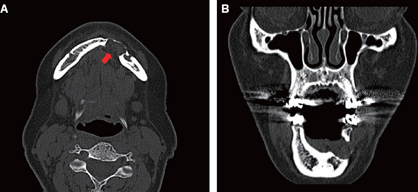
Fig. 2 (A) Computed tomography revealed an expansion of the buccal cortical bone of the left anterior mandible and perforation of the lingual bone (arrow) (horizontal view), (B) Computed tomography revealed penetration of the buccal and lingual bones of the left anterior mandible (coronal view).
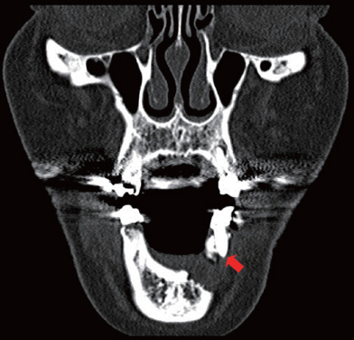
Fig. 3 Computed tomography detected no root resorption and normal configuration of root apex (arrow).
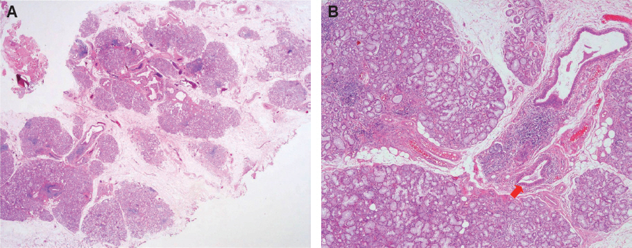
Fig. 4 Histological review showed lobules of seromucinous salivary gland including draining ducts (arrow) (H&E, (A) x10, (B) x40).
Reference
-
References
1. Kim H, Seok JY, Lee S, An J, Kim NR, Chung DH, Cho HY, Ha SY. Bilateral stafne bone cavity in the anterior mandible with heterotopic salivary gland tissue: a case report. Korean J Pathol. 2014; 48:248–9. DOI: 10.4132/KoreanJPathol.2014.48.3.248. PMID: 25013425. PMCID: PMC4087140.2. Probst FA, Probst M, Maistreli IZ, Otto S, Troeltzsch M. Imaging characteristics of a Stafne bone cavity-panoramic radiography, computed tomography and magnetic resonance imaging. Oral Maxillofac Surg. 2014; 18:351–3. DOI: 10.1007/s10006-014-0454-5. PMID: 25096915.3. Herranz-Aparicio J, Figueiredo R, Gay-Escoda C. Stafne’s bone cavity: an unusual case with involvement of the buccal and lingual mandibular plates. J Clin Exp Dent. 2014; 6:e96–9. DOI: 10.4317/jced.51229. PMID: 24596643. PMCID: PMC3935913.4. Fernandes M, de Ataide I. Nonsurgical management of periapical lesions. J Conserv Dent. 2010; 13:240–5. DOI: 10.4103/0972-0707.73384. PMID: 21217952. PMCID: PMC3010029.5. Katz J, Chaushu G, Rotstein I. Stafne’s bone cavity in the anterior mandible: a possible diagnostic challenge. J Endod. 2001; 27:304–7. DOI: 10.1097/00004770-200104000-00020. PMID: 11485274.6. Quesada-Gómez C, Valmaseda-Castellón E, Berini- Aytés L, Gay-Escoda C. Stafne bone cavity: a retrospective study of 11 cases. Med Oral Patol Oral Cir Bucal. 2006; 11:E277–80. PMID: 16648768.7. Prechtl C, Stockmann P, Neukam FW, Schlegel KA. Enlargement of a Stafne cyst as an indication for surgical treatment - a case report. J Craniomaxillofac Surg. 2013; 41:270–3. DOI: 10.1016/j.jcms.2012.10.013. PMID: 23218505.8. Boffano P, Gallesio C, Daniele D, Roccia F. An unusual trilobate Stafne bone cavity. Surg Radiol Anat. 2013; 35:351–3. DOI: 10.1007/s00276-012-1043-7. PMID: 23187427.9. Shimizu M, Osa N, Okamura K, Yoshiura K. CT analysis of the Stafne’s bone defects of the mandible. Dentomaxillofac Radiol. 2006; 35:95–102. DOI: 10.1259/dmfr/71115878. PMID: 16549436.10. Sisman Y, Etöz OA, Mavili E, Sahman H, Tarim Ertas E. Anterior Stafne bone defect mimicking a residual cyst: a case report. Dentomaxillofac Radiol. 2010; 39:124–6. DOI: 10.1259/dmfr/49320253. PMID: 20100926. PMCID: PMC3520195.11. Kopp S, Ihde S, Bienengraber V. Differential diagnosis of stafne idiopathic bone cyst with Digital Volume Tomography (DVT). J Maxillofac Oral Surg. 2010; 9:80–1. DOI: 10.1007/s12663-010-0023-x. PMID: 23139576. PMCID: PMC3453701.12. de Courten A, Küffer R, Samson J, Lombardi T. Anterior lingual mandibular salivary gland defect (Stafne defect) presenting as a residual cyst. Oral Surg Oral Med Oral Pathol Oral Radiol Endod. 2002; 94:460–4. DOI: 10.1067/moe.2002.125196. PMID: 12374920.13. Sisman Y, Miloglu O, Sekerci AE, Yilmaz AB, Demirtas O, Tokmak TT. Radiographic evaluation on prevalence of Stafne bone defect: a study from two centres in Turkey. Dentomaxillofac Radiol. 2012; 41:152–8. DOI: 10.1259/dmfr/10586700. PMID: 22074869. PMCID: PMC3520381.14. Branstetter BF, Weissman JL, Kaplan SB. Imaging of a Stafne bone cavity: what MR adds and why a new name is needed. AJNR Am J Neuroradiol. 1999; 20:587–9. PMID: 10319966.15. Ariji E, Fujiwara N, Tabata O, Nakayama E, Kanda S, Shiratsuchi Y, Oka M. Stafne’s bone cavity. Classification based on outline and content determined by computed tomography. Oral Surg Oral Med Oral Pathol. 1993; 76:375–80. DOI: 10.1016/0030-4220(93)90271-5.
- Full Text Links
-
- Actions
-
Cited
- CITED
-
- Close
- Share
-
- Similar articles
-
- Unusual Stafne bone cavity mimicking infected cyst or neural origin tumor
- A CASE REPORT: STAFNE'S CYST IN THE ANTERIOR MANDIBLE
- Comparison of digital radiometric featuresbetween radicular cysts and periapical granulomas
- Solitary Bone Cyst of the Proximal Femur Mimicking Fibrous Dysplasia: A Case Report
- A Case of Epidermoid Cyst at Nasolabial Area
