Healthc Inform Res.
2016 Jul;22(3):238-242. 10.4258/hir.2016.22.3.238.
Methods of Hematoxylin and Erosin Image Information Acquisition and Optimization in Confocal Microscopy
- Affiliations
-
- 1Biomedical Engineering Branch, Research Institute and Hospital, National Cancer Center, Goyang, Korea. gsgsbal@ncc.re.kr
- 2Molecular Imaging & Therapy Branch, Research Institute and Hospital, National Cancer Center, Goyang, Korea.
- 3Colorectal Cancer Branch, Research Institute and Hospital, National Cancer Center, Goyang, Korea.
- KMID: 2357402
- DOI: http://doi.org/10.4258/hir.2016.22.3.238
Abstract
OBJECTIVES
We produced hematoxylin and eosin (H&E) staining-like color images by using confocal laser scanning microscopy (CLSM), which can obtain the same or more information in comparison to conventional tissue staining.
METHODS
We improved images by using several image converting techniques, including morphological methods, color space conversion methods, and segmentation methods.
RESULTS
An image obtained after image processing showed coloring very similar to that in images produced by H&E staining, and it is advantageous to conduct analysis through fluorescent dye imaging and microscopy rather than analysis based on single microscopic imaging.
CONCLUSIONS
The colors used in CLSM are different from those seen in H&E staining, which is the method most widely used for pathologic diagnosis and is familiar to pathologists. Computer technology can facilitate the conversion of images by CLSM to be very similar to H&E staining images. We believe that the technique used in this study has great potential for application in clinical tissue analysis.
Keyword
MeSH Terms
Figure
-
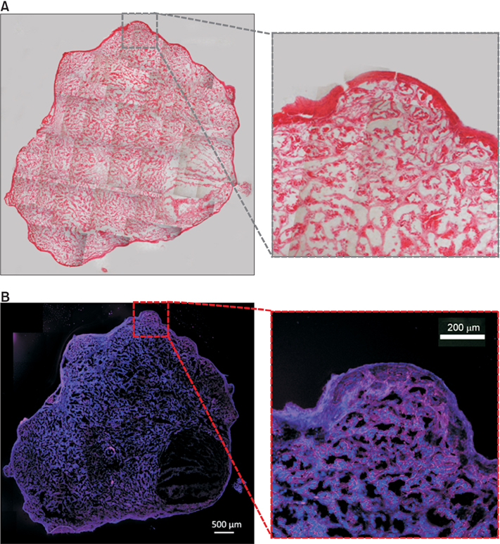
Figure 1 (A) Pictures of H&E stained tumor section, (B) fluorescence image of tumor section obtained using confocal laser scanning microscopy.
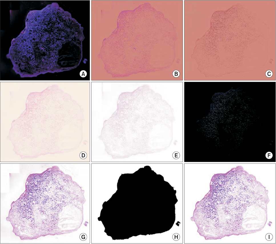
Figure 2 Images obtained during image processing procedure: (A) original fluorescence image, (B, C) conversion from RGB to CIE Luv, (D) merged images, (E) background image conversion, (F) nucleus creation image, (G) enhanced image, (H) mask creation image, and (I) final image.

Figure 3 Magnified image after the image processing procedure. Nuclei are blue, while cytoplasm and extracellular matrix are pink.
Reference
-
1. Esbona K, Li Z, Wilke LG. Intraoperative imprint cytology and frozen section pathology for margin assessment in breast conservation surgery: a systematic review. Ann Surg Oncol. 2012; 19(10):3236–3245.
Article2. Ferreiro JA, Myers JL, Bostwick DG. Accuracy of frozen section diagnosis in surgical pathology: review of a 1-year experience with 24,880 cases at Mayo Clinic Rochester. Mayo Clin Proc. 1995; 70(12):1137–1141.
Article3. Smith GJ, Bagnell CR, Bakewell WE, Black KA, Bouldin TW, Earnhardt TS, et al. Application of confocal scanning laser microscopy in experimental pathology. J Electron Microsc Tech. 1991; 18(1):38–49.
Article4. Fink-Puches R, Hofmann-Wellenhof R, Smolle J, Kerl H. Confocal laser scanning microscopy: a new optical microscopic technique for applications in pathology and dermatology. J Cutan Pathol. 1995; 22(3):252–259.
Article5. Ragazzi M, Piana S, Longo C, Castagnetti F, Foroni M, Ferrari G, et al. Fluorescence confocal microscopy for pathologists. Mod Pathol. 2014; 27(3):460–471.
Article6. Farkas DH. Diagnostic molecular pathology in an era of genomics and translational bioinformatics. Diagn Mol Pathol. 2008; 17(1):1–2.
Article7. De Rossi A, Rocha LB, Rossi MA. Application of fluorescence microscopy on hematoxylin and eosin-stained sections of healthy and diseased teeth and supporting structures. J Oral Pathol Med. 2007; 36(6):377–381.
Article8. Gareau D, Bar A, Snaveley N, Lee K, Chen N, Swanson N, et al. Tri-modal confocal mosaics detect residual invasive squamous cell carcinoma in Mohs surgical excisions. J Biomed Opt. 2012; 17(6):066018.
Article9. Portugal J, Waring MJ. Assignment of DNA binding sites for 4',6-diamidine-2-phenylindole and bisbenzimide (Hoechst 33258): a comparative footprinting study. Biochim Biophys Acta. 1988; 949(2):158–168.
Article10. Bradski G, Kaehler A. Learning OpenCV: computer vision with the OpenCV library. Sebastopol (CA): O'Reilly Media;2008.11. Velde KV. Multi-scale color image enhancement. In : Proceedings of 1999 International Conference on Image Processing (ICIP); 1999 Oct 24-28; Kobe, Japan. p. 584–587.12. Kruse FA, Raines GL. Technique for enhancing digital color images by contrast stretching in munsell color space. In : Proceedings of the International Symposium on Remote Sensing of Environment, Third Thematic Conference: Remote Sensing for Exploration Geology; 1984 Apr 16-19; Colorado Springs, CO. p. 755–760.
- Full Text Links
-
- Actions
-
Cited
- CITED
-
- Close
- Share
-
- Similar articles
-
- Correction: Methods of Hematoxylin and Eosin Image Information Acquisition and Optimization in Confocal Microscopy
- Applications of Artificial Intelligence in MR Image Acquisition and Reconstruction
- Comparision of Specular Microscopy and Confocal Microscopy for Evaluation of Corneal Endothelium
- In vivo tandem scanning confocal microscopy in acanthamoeba keratitis
- Optimization of Subtraction Brain Perfusion SPECT with Basal / Acetazolamide Consecutive Acquisition
