J Korean Soc Med Inform.
2009 Mar;15(1):165-172.
Detection of Microcalcifications in Digital Mammograms Using Foveal Method
- Affiliations
-
- 1Biomedical Engineering Branch, Division of Cancer Biology, National Cancer Center, Korea.
- 2Center for Breast cancer, National Cancer Center, Korea.
- 3Breast & Endocrine Cancer Branch, National Cancer Center, Korea.
- 4Diagnostic Radiology, Seoul National University Hospital, Seoul National University, Korea.
Abstract
OBJECTIVE
Breast cancer represents themost frequently diagnosed cancer in women. In order to reduce mortality, early detection of breast cancer is important, because diagnosis is more likely to be successful in the early stages of the disease. On the average, the reader's sensitivity can be increased by 10%with the assistance of computer-aided diagnosis (CAD) system. This paper presents a CAD system for the automatic detection of clustered micro-calcifications in digitized mammograms.
METHODS
The proposed system consists of three main steps. First, breast region is segmented from original mammogram using contrast property of grey level co-occurrence matrix(GLCM). Second, potential micro-calcification pixels in the mammograms are detected by foveal method. Third, in order to reduce false-positive rate, individual micro-calcifications are detected by a set of 8 features extracted from the potential individual micro-calcification objects.
RESULTS
In the result, Specificity and sensitivity are used to evaluate the detection performance of micro-calcifications.(sensitivity : 93.1%, specificity : 87.5%).
CONCLUSION
This study could be a useful method for diagnosis of breast cancer as a CAD system.
Keyword
Figure
-

Figure 1 An example of micro-calcification in mammogram image
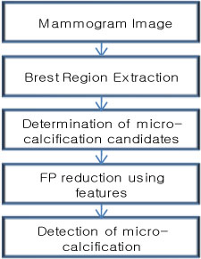
Figure 2 The flow chart of our CAD system for detection of micro-calcification.

Figure 3 The result of breast region segmentation
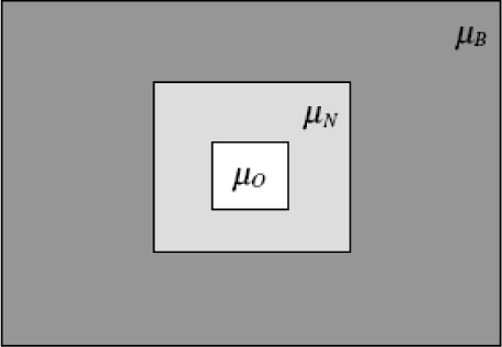
Figure 4 The foveal masks used for the computation of µ0, µN and µB. The O is the size of the kernel object, N its neighborhood and B the background
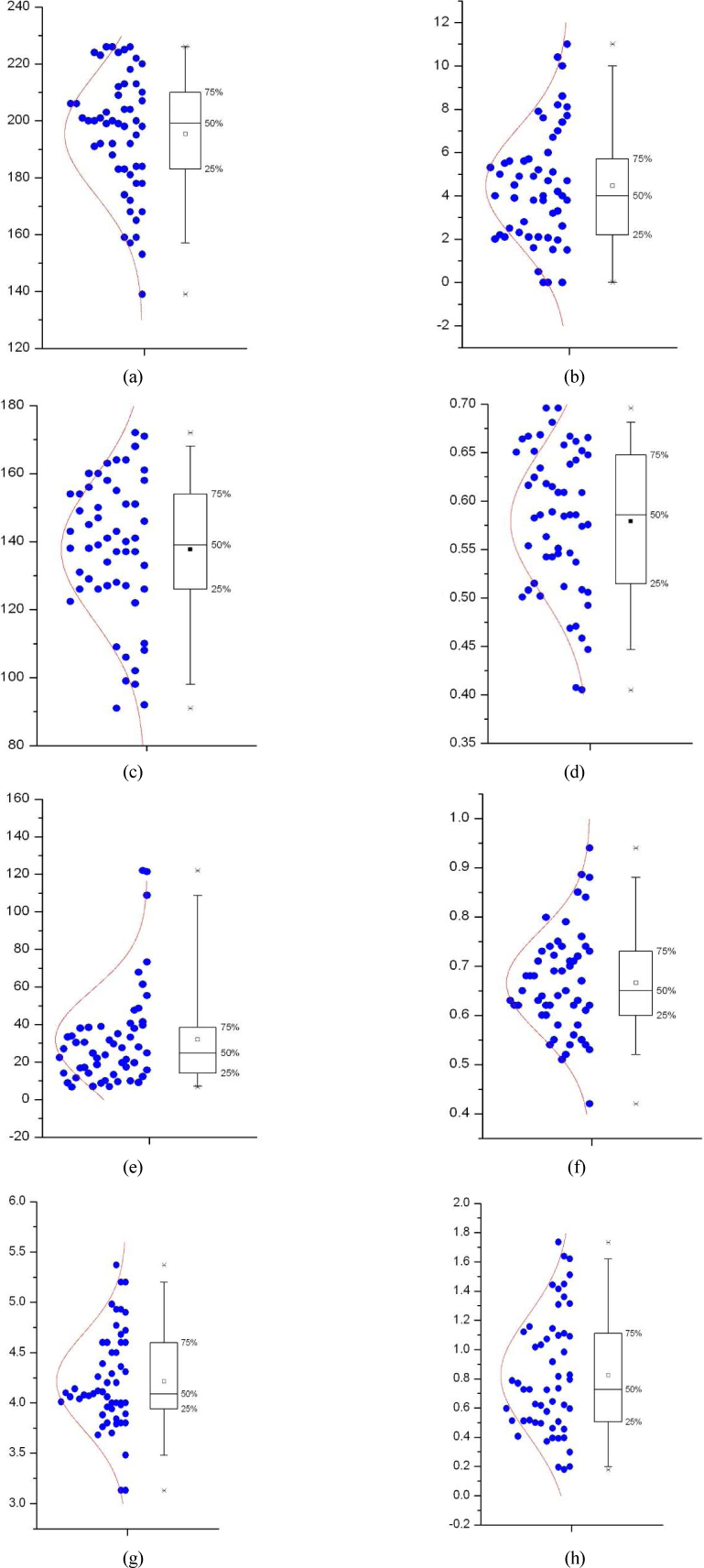
Figure 5 (a) Mean (b) Standard deviation (c) Foreground background difference (d) Difference ratio (e) GLCM contrast (f) GLCM correlation (g) GLCM entropy (h) Fractal dimension hurst coefficient

Figure 6 The result image of proposed method (a),(c) original image, (b),(d) result image
Reference
-
1. Feig S.A. Decreased breast cancer mortality through mammographic screening : Results of a clinical trial. Radiology. 1988. 167(3):659–665.
Article2. Dhawan A.P., Le Royer E. Mammographic feature enhancement by computerized image processing. Comput Methods Programs Biomed. 1988. 27(1):23–25.
Article3. Chan H.P., Doi K., Galhotra S., Vyborny C.J., MacMahon H., Jokich P.M. Image feature analysis and computer-aided diagnosis in digital radiography I. Automated detection of microcalcifications in mammography. Med Phys. 1987. 14(4):538–548.
Article4. Chan H.P., Doi K., Vyborny C.J., Lam K.L., Schmidt R.A. Computer-aided detection of microcalcifications in mammograms-methodology and preliminary calinical study. Invest Radiol. 1988. 23(9):664–671.5. Olson S.L., Fam B.W., Winter P.F., Scholz F.J., Lee A.K., Gordon S.E. Breast calcifications:analysis of imaging properties. Radiology. 1988. 169(2):329–332.
Article6. Sickles E.A. Mammographic features of 300 consecutive nonpalpable breast cancers. Am J Radiology. 1986. 146:661–665.
Article7. Nishikawa R.M. Detection of microcalcification. Digital Mammography Nijmegen 98. 1998. Amsterdam: Kluwer Academic Publisher;131–153.8. Davies D.H., Dance D.R. The automatic computer detection of subtle calcification in radiographically dense breasts. Phys Med Biol. 1992. 37:1385–1390.
Article9. Zhang W., Doi K., Giger M.L., Wu Y., Nishikawa R.M., Schmidt R.A. Computerized detection of clustered microcalcifications in digital mammograms using a shift-invariant artificial neural network. Med Phys. 1994. 21:517–524.
Article10. Wu Y., Doi K., Giger M.L., Nishikawa R.M. Computerized detection of clustered microcalcifiations in digital mammograms:Applications of artificial neural networks. Med Phys. 1992. 19:555–560.
Article11. Karssemeijer N. Adaptive noise equalisation and recognition of microcalcification clusters in mammograms. International Journal of Pattern Recognition and Artificial Intelligence. 1993. 7(6):1357–1372.12. Chan H.P, et al. Improvement in radiologists' detection of clustered microcalcifications on mammograms. The potential of computer-aided diagnosis. Invest Radiol. 1990. 25(10):1102–1110.
Article13. Chan H.P, et al. Computer-aided detection of microcalcifications in mammograms. Methodology and preliminary clinical study. Invest Radiol. 1988. 23(9):664–671.14. Zhang W, et al. Optimallyweightedwavelet transform based on supervised training for detection of microcalcifications in digital mammograms. Med Phys. 1998. 25(6):949–956.
Article15. Shen L., Rangayyan R.M., Desautels J.L. Application of shape analysis to mammographic calcifications. IEEE Trans Med Imaging. 1994. 13(2):263–274.
Article16. Heucke L., Knaak M., Orglmeister R. A newimage segmentation method based on human brightness perception and foveal adaptation. IEEE Signal Processing Letters. 2000. 7(6):129–131.
Article17. Highnam R.P., Brady J.M. Mammographic Image Analysis. 1999. London: Kluwer Academic Publishers.18. Kim M.S., Lee S.B. Design & Development of 3D Medical Image Processing System using VTK. Journal of Korean Society of Medical Informatics. 2003. 9(4):375–380.
Article
- Full Text Links
-
- Actions
-
Cited
- CITED
-
- Close
- Share
-
- Similar articles
-
- Reproducibility of Computer-Aided Detection Marks in Digital Mammography
- Screening Mammography-Detected Cancers: The Sensitivity of the Computer-aided Detection System as Applied to Full-field Digital Mammography
- Pathologic Analysis of Clustered Microcalcification Found on Mammograms: A Review of 77 Cases
- Breast Cancer Incidence According to Microcalcification Types on Mammogram
- Hyperdense Dots Mimicking Microcalcifications: Mammographic Findings
