Overexpression of Endoplasmic Reticulum Oxidoreductin 1-α (ERO1L) Is Associated with Poor Prognosis of Gastric Cancer
- Affiliations
-
- 1Department of Medical Oncology, Gangnam Severance Cancer Hospital, Yonsei University College of Medicine, Seoul, Korea. chojy@yuhs.ac
- 2Samsung Bioepis Co., Ltd., Incheon, Korea.
- 3Department of Pathology, Gangnam Severance Cancer Hospital, Yonsei University College of Medicine, Seoul, Korea.
- 4Department of Surgery, Gangnam Severance Cancer Hospital, Yonsei University College of Medicine, Seoul, Korea.
- KMID: 2356222
- DOI: http://doi.org/10.4143/crt.2015.189
Abstract
- PURPOSE
Gastric cancer is the second leading cause of cancer-related death worldwide. Although surgery is the standard curative treatment for gastric cancer, relapse occurs in a large number of patients, except in the case of early diagnosed gastric cancer. Following previous studies that identified endoplasmic reticulum oxidoreductin 1-α (ERO1L) as a potential marker for gastric cancer, we investigated the functional role of ERO1L in gastric cancer.
MATERIALS AND METHODS
For validation of microarray data, the mRNA expression level of ERO1L was measured by quantitative real-time reverse transcription polymerase chain reaction in 56 independent stage III gastric cancer patients. Immunohistochemical staining was performed to examine the protein expression level of ERO1L in 231 gastric cancer patients. Correlation between gene expression and cancer prognosis was evaluated.
RESULTS
Patients with high ERO1L expression had poorer survival than those with low expression (p < 0.01). Functional assays demonstrated that ERO1L knockdown inhibited cell proliferation, migration, invasion, and chemoresistance. In addition, involvement of inactivation of Akt and JNK signaling in molecular mechanisms of ERO1L inhibition was demonstrated.
CONCLUSION
High expression of ERO1L is associated with poor prognosis of patients with gastric cancer. These results indicate that ERO1L expression may be a clinically promising therapeutic target for prevention of gastric cancer.
MeSH Terms
Figure
-
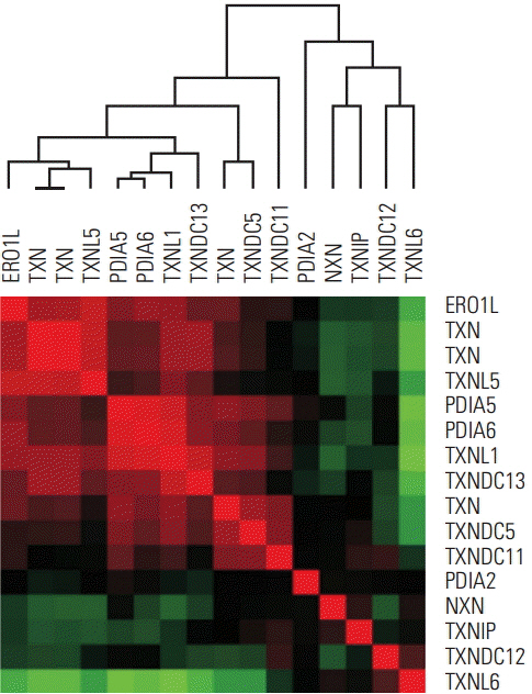
Fig. 1. ERO1L and TXN family in-trans correlation in human gastric cancer. Data are given in NCBI’s GEO public database (microarray data accession number, GSE13861). The color red or green reflects relative high or low expression level, respectively. ERO1L, endoplasmic reticulum oxidoreduction 1-α; TXN, thioredoxin.
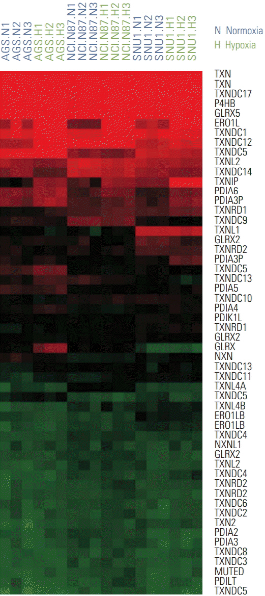
Fig. 2. Two different conditions of gastric cancer cell lines, normoxia and hypoxia, show mRNA expression patterns. mRNA expression patterns of gastric cancer cells (AGS, NCI-N87, and SNU1) in normoxia and hypoxia are depicted in a heat map. The color red or green reflects relative high or low expression level, respectively.
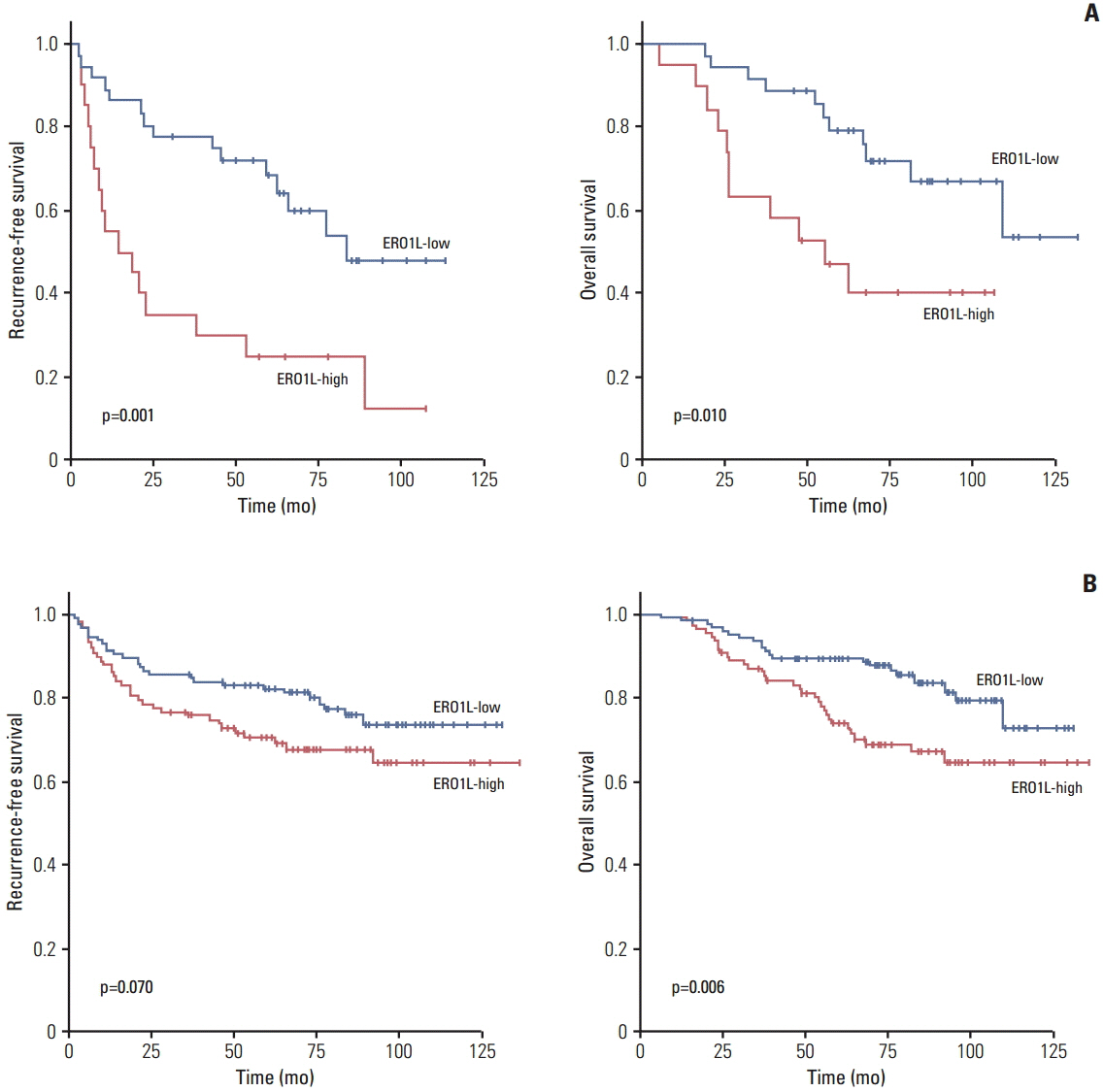
Fig. 3. Kaplan-Meier plot of recurrence-free survival (RFS) and overall survival (OS) according to gene expression. (A) Gene expression was measured using quantitative reverse transcription polymerase chain reaction in 56 stage III gastric cancer patients. (B) Gene expression was measured using immunohistochemical staining analysis in 231 gastric cancer patients divided into two groups according to their gene expression levels. ERO1L, endoplasmic reticulum oxidoreduction 1-α.

Fig. 4. Representative images of endoplasmic reticulum oxidoreduction 1-α (ERO1L) protein expression in gastric cancer. High immunohistochemical staining of ERO1L in cytoplasm of gastric cancer (left, ×40; middle, ×100; right, ×200).

Fig. 5. Endoplasmic reticulum oxidoreduction 1-α (ERO1L) expression in gastric cancer cell lines. (A) ERO1L mRNA levels were assessed using quantitative real-time reverse transcription polymerase chain reaction. Expression of β-actin was included as an internal loading control. (B) ERO1L protein levels were analyzed using immunoblotting. Glyceraldehyde 3-phosphate dehydrogenase (GAPDH) was included as an internal loading control.
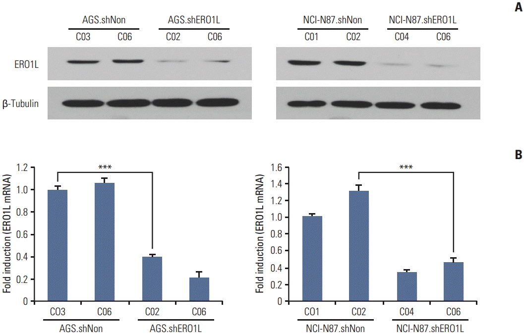
Fig. 6. Endoplasmic reticulum oxidoreduction 1-α (ERO1L) knockdown expression in lentivirus-mediated stable cells. Total RNA and whole cell lysates were collected from AGS and NCI-N87 cells transfected with shNonTarget and shERO1L vector (AGS, shNon.C03 and C06/shERO1L.C02 and C06; NCI-N87, shNon.C01 and C02/shERO1L.C04 and C06). (A) Expression of ERO1L protein was analyzed by immunoblotting, and mRNA level was measured by quantitative real-time reverse transcription polymerase chain reaction. β-Tubulin and β-actin were included as an internal loading control. ***p < 0.001.

Fig. 7. Endoplasmic reticulum oxidoreduction 1-α (ERO1L)–deficient gastric cancer cells decrease cell proliferation. (A) Cell proliferation was determined by WST-1 assay. Cell proliferation curves for AGS and NCI-N87 cells at indicated times. Error bars represent mean±standard deviation (SD) of triplicate experiments. (B) A clonogenic assay was performed on AGS and NCI-N87 cells for 21 days. Left panel shows representative images, and right panel shows quantification of colonies. Error bars represent mean±standard deviation of triplicate experiments. ***p < 0.001.
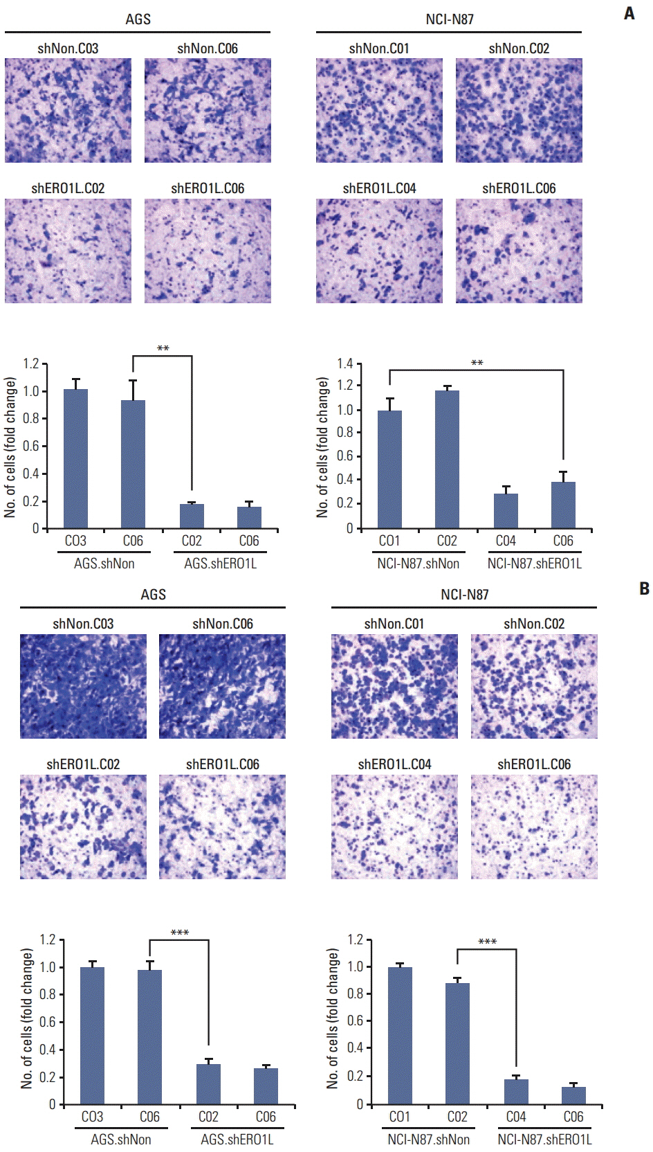
Fig. 8. Silencing of endoplasmic reticulum oxidoreduction 1-α(ERO1L) expression inhibits migration and invasion abilities of AGS and NCI-N87 cells. (A) Cell migration was determined using Boyden chambers. (B) Cell invasion was assayed using a membrane coated with Matrigel. Five random microscopic fields were counted for each group. The results presented are an average of five random microscopic fields from three independent experiments. **p < 0.01, ***p < 0.001.
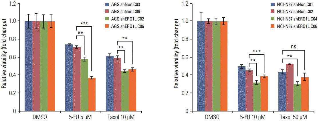
Fig. 9. Endoplasmic reticulum oxidoreduction 1-α (ERO1L)–deficient gastric cancer cells reduce chemoresistance. Viability of ERO1L-deficient gastric cancer cells treated for 48 hours with chemotherapeutic agents (paclitaxel and 5-fluorouracil [5-FU]) measured by WST-1 assay. Cells treated with dimethylsulfoxide (DMSO) as a control. Data are shown as relative viability (fold change) to the control groups. Data are mean±standard deviation of triplicate experiments. **p < 0.01, ***p < 0.001; ns, not significant.
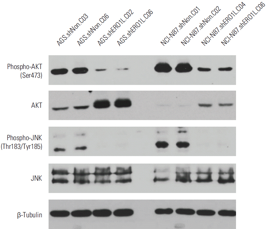
Fig. 10. Inhibitory effects of endoplasmic reticulum oxidoreduction 1-α (ERO1L)–deficient gastric cancer cells on Akt and JNK signaling pathway. Expression levels of total Akt, phosphorylated Akt at Ser473, total JNK, phosphorylated JNK at Thr183/Tyr185, and β-tubulin (internal control) were measured in the cell lysates. Akt phosphorylation at Ser473 and JNK phosphorylation at Thr183/Tyr185 surprisingly decreased in ERO1L-deficient gastric cancer cells (AGS, shERO1L.C02 and C06; NCI-N87, shERO1L.C04 and C06).
Cited by 1 articles
-
Increased Progastrin-Releasing Peptide Expression is Associated with Progression in Gastric Cancer Patients
Li Li, Xiaodong Yin, Hai Meng, Juanyu Hu, Zhengqing Yu, Jianyong Xu
Yonsei Med J. 2020;61(1):15-19. doi: 10.3349/ymj.2020.61.1.15.
Reference
-
References
1. Jemal A, Bray F, Center MM, Ferlay J, Ward E, Forman D. Global cancer statistics. CA Cancer J Clin. 2011; 61:69–90.
Article2. Lynch HT, Grady W, Suriano G, Huntsman D. Gastric cancer: new genetic developments. J Surg Oncol. 2005; 90:114–33.
Article3. Shah MA, Ajani JA. Gastric cancer: an enigmatic and heterogeneous disease. JAMA. 2010; 303:1753–4.4. Gallo A, Cha C. Updates on esophageal and gastric cancers. World J Gastroenterol. 2006; 12:3237–42.
Article5. Bang YJ, Kim YW, Yang HK, Chung HC, Park YK, Lee KH, et al. Adjuvant capecitabine and oxaliplatin for gastric cancer after D2 gastrectomy (CLASSIC): a phase 3 open-label, randomised controlled trial. Lancet. 2012; 379:315–21.
Article6. Tay ST, Leong SH, Yu K, Aggarwal A, Tan SY, Lee CH, et al. A combined comparative genomic hybridization and expression microarray analysis of gastric cancer reveals novel molecular subtypes. Cancer Res. 2003; 63:3309–16.7. Kim B, Bang S, Lee S, Kim S, Jung Y, Lee C, et al. Expression profiling and subtype-specific expression of stomach cancer. Cancer Res. 2003; 63:8248–55.8. Chen X, Leung SY, Yuen ST, Chu KM, Ji J, Li R, et al. Variation in gene expression patterns in human gastric cancers. Mol Biol Cell. 2003; 14:3208–15.
Article9. Lee HS, Cho SB, Lee HE, Kim MA, Kim JH, Park DJ, et al. Protein expression profiling and molecular classification of gastric cancer by the tissue array method. Clin Cancer Res. 2007; 13:4154–63.
Article10. Tan IB, Ivanova T, Lim KH, Ong CW, Deng N, Lee J, et al. Intrinsic subtypes of gastric cancer, based on gene expression pattern, predict survival and respond differently to chemotherapy. Gastroenterology. 2011; 141:476–85.
Article11. Ooi CH, Ivanova T, Wu J, Lee M, Tan IB, Tao J, et al. Oncogenic pathway combinations predict clinical prognosis in gastric cancer. PLoS Genet. 2009; 5:e1000676.
Article12. Cho JY, Lim JY, Cheong JH, Park YY, Yoon SL, Kim SM, et al. Gene expression signature-based prognostic risk score in gastric cancer. Clin Cancer Res. 2011; 17:1850–7.
Article13. Bristow RG, Hill RP. Hypoxia and metabolism: hypoxia, DNA repair and genetic instability. Nat Rev Cancer. 2008; 8:180–92.14. Benham AM, Cabibbo A, Fassio A, Bulleid N, Sitia R, Braakman I. The CXXCXXC motif determines the folding, structure and stability of human Ero1-Lalpha. EMBO J. 2000; 19:4493–502.
Article15. Gess B, Hofbauer KH, Wenger RH, Lohaus C, Meyer HE, Kurtz A. The cellular oxygen tension regulates expression of the endoplasmic oxidoreductase ERO1-Lalpha. Eur J Biochem. 2003; 270:2228–35.16. May D, Itin A, Gal O, Kalinski H, Feinstein E, Keshet E. Ero1-L alpha plays a key role in a HIF-1-mediated pathway to improve disulfide bond formation and VEGF secretion under hypoxia: implication for cancer. Oncogene. 2005; 24:1011–20.17. Simon R, Lam A, Li MC, Ngan M, Menenzes S, Zhao Y. Analysis of gene expression data using BRB-ArrayTools. Cancer Inform. 2007; 3:11–7.18. Eisen MB, Spellman PT, Brown PO, Botstein D. Cluster analysis and display of genome-wide expression patterns. Proc Natl Acad Sci U S A. 1998; 95:14863–8.
Article19. Bellacosa A, Kumar CC, Di Cristofano A, Testa JR. Activation of AKT kinases in cancer: implications for therapeutic targeting. Adv Cancer Res. 2005; 94:29–86.
Article20. Malik SN, Brattain M, Ghosh PM, Troyer DA, Prihoda T, Bedolla R, et al. Immunohistochemical demonstration of phospho-Akt in high Gleason grade prostate cancer. Clin Cancer Res. 2002; 8:1168–71.21. Kreisberg JI, Malik SN, Prihoda TJ, Bedolla RG, Troyer DA, Kreisberg S, et al. Phosphorylation of Akt (Ser473) is an excellent predictor of poor clinical outcome in prostate cancer. Cancer Res. 2004; 64:5232–6.
Article22. Ali A, Sidorova TS, Matesic DF. Dual modulation of JNK and Akt signaling pathways by chaetoglobosin K in human lung carcinoma and ras-transformed epithelial cells. Invest New Drugs. 2013; 31:525–34.
Article23. Engelberg D. Stress-activated protein kinases-tumor suppressors or tumor initiators? Semin Cancer Biol. 2004; 14:271–82.
Article
- Full Text Links
-
- Actions
-
Cited
- CITED
-
- Close
- Share
-
- Similar articles
-
- Endoplasmic Reticulum Stress and Diabetes
- New Insights into the Role of Endoplasmic Reticulum Stress in Breast Cancer Metastasis
- Endoplasmic Reticulum (ER) Stress and Vascular Complication
- Clinicopathological Features and Prognosis of Gastric Cancer in Young Patients
- Nuclear Receptors Resolve Endoplasmic Reticulum Stress to Improve Hepatic Insulin Resistance
