Fusiform Superior Cerebellar Artery Aneurysm Treated with Endovascular Treatment
- Affiliations
-
- 1Department of Neurosurgery, Inha University School of Medicine and Hospital, Incheon, Korea. dro3@nate.com
- KMID: 2355654
- DOI: http://doi.org/10.7461/jcen.2016.18.3.276
Abstract
- An aneurysm of the distal superior cerebellar artery (SCA) is a highly rare disease. Fusiform aneurysms of the distal SCA are particularly challenging to treat. Clipping, trapping with or without bypass using microsurgery or endovascular treatment (EVT) were used to treat this condition. We describe the case of fusiform distal SCA aneurysms treated successfully with endovascular coiling with a 3-month follow-up. A 39 year-old male was presented with subarachnoid hemorrhage (SAH) and a 15 mm fusiform aneurysm of the ambient segment of the left distal SCA. EVT for parent artery occlusion and packing of the aneurysm was done. Left sixth nerve palsy appeared after 1 day of EVT. The symptom completely recovered within 1 week of the post-procedural period. No neurological deficit was seen during the clinical 3-month follow-up. EVT of fusiform distal SCA aneurysms with coils is a safe and feasible option to manage this rare condition. However, the treatment options must be carefully selected depending on the neurologic condition, development of collateral circulation, and configuration of the dissection.
MeSH Terms
Figure
-
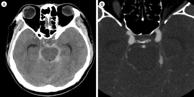
Fig. 1 (A) Brain computed tomography (CT) image. The brain CT shows a Fisher Grade III SAH in the pre-pontine, left ambient and quadrigeminal cistern. (B) Brain computed tomography angiography (CT angiography) image. The brain CT angiography shows fusiform dilatation of the left distal SCA. SAH = subarachnoid hemorrhage; SCA = superior cerebellar artery.
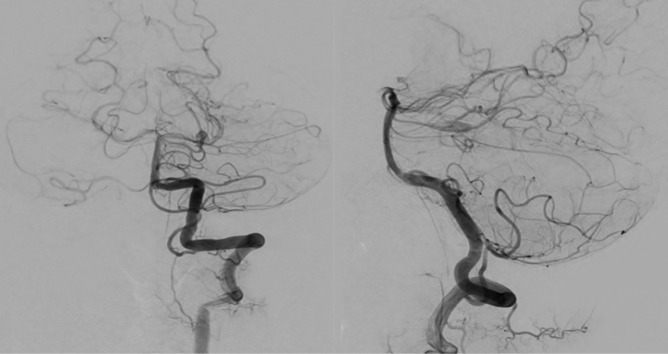
Fig. 2 Digital subtraction angiography (DSA) images. The DSA images show the fusiform aneurysm of the left superior cerebellar artery. There was sufficient collateral circulation from the ipsilateral AICA and PICA. AICA = anterior inferior cerebellar artery; PICA = posterior inferior cerebellar artery.
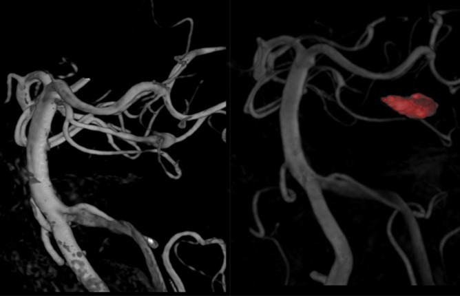
Fig. 3 Pre-operative 3-dimensional (D) CT angiography (left) and post-operative 3D DSA images (right). The 3D CT angiography reveals fusiform dilatation of the left distal SCA (left). The 3D DSA shows complete occlusion of the aneurysm sac and involved the distal SCA branch. The ipsilateral duplicated SCA is intact. CT = computed tomography; DSA = digital subtraction angiography; SCA = superior cerebellar artery.
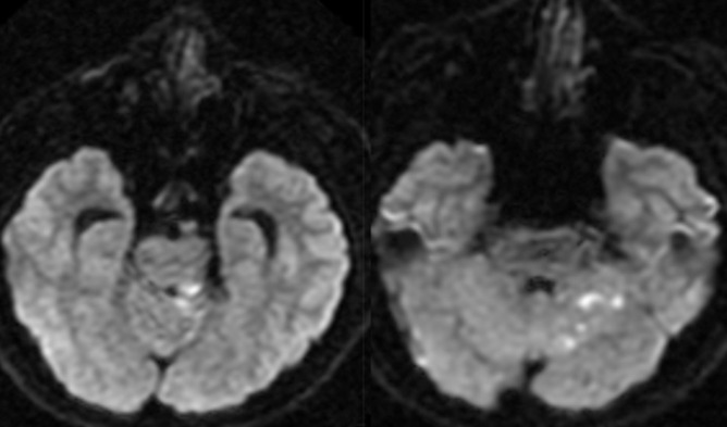
Fig. 4 Diffusion weighted images (DWIs) 1 day post-treatment. The DWIs show scattered high signal change, suggesting acute infarction on the left posterior tegmentum, superior vermis, and superior part of the left cerebellum.
Reference
-
1. Alurkar A, Karanam LSP, Nayak S, Oak S. Endovascular management of fusiform superior cerebellar artery aneurysms: a series of three cases with review of literature. J Clin Imaging Sci. 2012; 7. 2:47. PMID: 22919561.
Article2. Biondi A. Trunkal intracranial aneurysms: dissecting and fusiform aneurysms. Neuroimaging Clin N Am. 2006; 8. 16(3):453–465. viiiPMID: 16935710.
Article3. Bozboğa M, Canbolat A, Savaş A, Türker K. Aneurysm arising from the medial branch of the superior cerebellar artery. Acta Neurochir (Wien). 1996; 8. 138(8):1013–1014. PMID: 8891001.
Article4. Briganti F, Marseglia M, Leone G, Briganti G, Piccolo D, Napoli M, et al. Endovascular treatment of a small aneurysm of the superior cerebellar artery with a flow-diverter device. A case report. Neuroradiol J. 2013; 6. 26(3):327–331. PMID: 23859291.5. Chaloupka JC, Putman CM, Awad IA. Endovascular therapeutic approach to peripheral aneurysms of the superior cerebellar artery. AJNR Am J Neuroradiol. 1996; 8. 17(7):1338–1342. PMID: 8871721.6. Chang SW, Abla AA, Kakarla UK, Sauvageau E, Dashti SR, Nakaji P, et al. Treatment of distal posterior cerebral artery aneurysms: a critical appraisal of the occipital artery-to-posterior cerebral artery bypass. Neurosurgery. 2010; 7. 67(1):16–25. discussion 25-6. PMID: 20559088.7. Danet M, Raymond J, Roy D. Distal superior cerebellar artery aneurysm presenting with cerebellar infarction: report of two cases. AJNR Am J Neuroradiol. 2001; 4. 22(4):717–720. PMID: 11290485.8. Haruma J, Sugiu K, Shimazu Y, Michiue H, Tokunaga K, Date I. Surgical and endovascular treatment for superior cerebellar artery aneurysms: report of two cases. No Shinkei Geka. 2013; 1. 41(1):45–51. PMID: 23269255.9. Ikeda K, Shoin K, Taguchi H, Yamano J, Kawahara R. Postpartum dissecting aneurysm of the superior cerebellar artery--case report. Neurol Med Chir (Tokyo). 1999; 11. 39(12):852–857. PMID: 10639812.
- Full Text Links
-
- Actions
-
Cited
- CITED
-
- Close
- Share
-
- Similar articles
-
- Successful Endovascular Treatment of Ruptured Superior Cerebellar Artery Aneurysm Associated with Moyamoya Disease : A Case Report and Review of the Literature
- Coil Embolization of Ruptured Thrombosed Distal Superior Cerebellar Artery Aneurysm: A Case Report
- Giant fusiform aneurysm at the basilar trunk treated with endovascular coil occlusion following bypass surgery for the flow diversion
- Stent-Assisted Coil Trapping in a Manual Internal Carotid Artery Compression Test for the Treatment of a Fusiform Dissecting Aneurysm
- Endovascular Treatment of a Fusiform Aneurysm Involving a Premammillary Artery Originating from the Internal Carotid Artery: A Case Report
