Clinical Utility of Angiographic CT with a Flat-detector Angiographic System during Endovascular Procedure
- Affiliations
-
- 1Department of Neurosurgery, Bucheon St Mary's Hospital, College of Medicine, The Catholic University of Korea, Bucheon, Korea. armada1997@naver.com
- KMID: 2355649
- DOI: http://doi.org/10.7461/jcen.2016.18.3.247
Abstract
OBJECTIVE
We evaluated the feasibility of angiographic computed tomography (ACT) for visualizing stent material in patients who underwent intracranial or extracranial stent placement to treat atherosclerotic lesions or stent assisted coil embolization.
MATERIALS AND METHODS
We performed intrarterial and intravenous ACT on biplane angiography system equipped with flat panel detectors (Axiom Arits dBA; Siemens Medical Solutions, Forchheim, Germany). Vistipaque 320 was injected for contrast medium, total 150 mL at flow rate of 5 mL/s through artery and 77 mL at flow rate of 3.5 mL/s through vein.
RESULTS
ACT is a new imaging modality that provides a clear visualization of stent strut.
CONCLUSION
Therefore this new application has potential to become the noninvasive option for follow-up after endovascular surgery using stents.
MeSH Terms
Figure
-
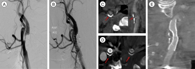
Fig. 1 (A) Stenosis, 85% (B) Precise 8 × 40 mm stent insertion. (C) Preop IA-ACT shows calcified plaque (arrow). (D) On axial views on post op IA and IV-ACT, the level of the plaque are showen. They further reveal the circumferential narrowing by calcified plaque (arrow). (E) Lateral ACT view (1 mm sections) provide superior visualization of the entire stent its strus and the remanining narrowing and show the underlying, heavialy calcified plaque(arrowhead) responsible for incomplete stent deployment. IA = intrarterial; ACT = angiographic computed tomography; IV = intravenous.
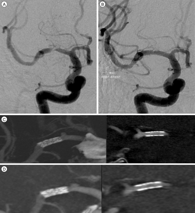
Fig. 2 (A) Severe stenosis of the right M1 segment. (B) Treatment with 2.5 × 8 mm durg eluting stent (Endeavor; Medtronic scientific, Roncadelle (BS), Italy) with successful, completely reestablishing the lumen (C) IA-ACT immediately after procedure show clear visualization of the stent and distal patency, also show no acute thrombosis. (D) IV-ACT after 3 month later show no instent stenois and clear stent struct. IA = intrarterial; ACT = angiographic computed tomography; IV = intravenous.
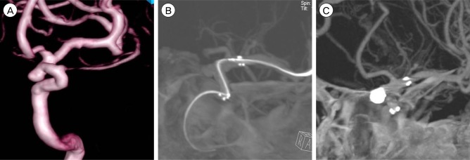
Fig. 3 (A) 3D DSA show unruptured aneurysm on paraclinoid aneurysm. (B) IA-ACT is performed after stent deployment and before coil embolizatoin. Excellent stent distal and proximal marker visibility and good position of stent is shown. (C) ACT image is performed after coil embolization allows definite exclusion of stent strut movement or coil protrusion with minimal beam-hardening artifacts. DSA = digital subtraction angiography; ACT = angiographic computed tomography.
Reference
-
1. Benitez RP, Silva MT, Klem J, Veznedaroglu E, Rosenwasser RH. Endovascular occlusion of wide-necked aneurysms with a new intracranial microstent (Neuroform) and detachable coils. Neurosurgery. 2004; 6. 54(6):1359–1367. discussion 1368. PMID: 15157292.
Article2. Benndorf G, Claus B, Strother CM, Chang L, Klucznik RP. Increased cell opening and prolapse of struts of a neuroform stent in curved vasculature: value of angiographic computed tomography: technical case report. Neurosurgery. 2006; 4. 58(4 Suppl 2):ONS-E380. discussion ONS-E380.3. Benndorf G, Strother CM, Claus B, Naeini R, Morsi H, Klucznik R, et al. Angiographic CT in cerebrovascular stenting. AJNR Am J Neuroradiol. 2005; 8. 26(7):1813–1818. PMID: 16091535.4. Buhk JH, Lingor P, Knauth M. Angiographic CT with intravenous administration of contrast medium is a noninvasive option for follow-up after intracranial stenting. Neuroradiology. 2008; 4. 50(4):349–354. PMID: 18246336.
Article5. Grzyska U, Freitag J, Zeumer H. Selective cerebral intraarterial DSA. Complication rate and control of risk factors. Neuroradiology. 1990; 32(4):296–299. PMID: 2234388.6. Gupta R, Grasruck M, Suess C, Bartling SH, Schmidt B, Stierstorfer K, et al. Ultra-high resolution flat-panel volume CT: fundamental principles, design architecture, and system characterization. Eur Radiol. 2006; 6. 16(6):1191–1205. PMID: 16528556.
Article7. Howington JU, Hanel RA, Harrigan MR, Levy EI, Guterman LR, Hopkins LN. The Neuroform stent, the first microcatheter-delivered stent for use in the intracranial circulation. Neurosurgery. 2004; 1. 54(1):2–5. PMID: 14683535.
Article8. Jiang WJ, Xu XT, Du B, Dong KH, Jin M, Wang QH, et al. Long-term outcome of elective stenting for symptomatic intracranial vertebrobasilar stenosis. Neurology. 2007; 3. 68(11):856–858. PMID: 17353474.
Article9. Lee YJ, Kim DJ, Suh SH, Lee SK, Kim J, Kim DI. Stent-assisted coil embolization of intracranial wide-necked aneurysms. Neuroradiology. 2005; 9. 47(9):680–689. PMID: 16028036.
Article10. Schueler BA, Kallmes DF, Cloft HJ. 3D cerebral angiography: radiation dose comparison with digital subtraction angiography. AJNR Am J Neuroradiol. 2005; 9. 26(8):1898–1901. PMID: 16155131.11. SSYLVIA Study Investigators. Stenting of Symptomatic Atherosclerotic Lesions in the Vertebral or Intracranial Arteries (SSYLVIA): study results. Stroke. 2004; 6. 35(6):1388–1392. PMID: 15105508.12. Trossbach M, Hartmann M, Braun C, Sartor K, Hähnel S. Small vessel stents for intracranial angioplasty: in vitro evaluation of in-stent stenoses using CT angiography. Neuroradiology. 2004; 6. 46(6):459–463. PMID: 15127168.
Article13. Wanke I, Doerfler A, Goericke S, Gizewski ER, Sandalcioglu E, Moemken S, et al. Treatment of wide-necked intracranial aneurysms with a self-expanding stent: mid-term results. Zentralbl Neurochir. 2005; 11. 66(4):163–169. PMID: 16317598.
Article14. Wanke I, Doerfler A, Schoch B, Stolke D, Forsting M. Treatment of wide-necked intracranial aneurysms with a self-expanding stent system: initial clinical experience. AJNR Am J Neuroradiol. 2003; Jun-Jul. 24(6):1192–1199. PMID: 12812954.
- Full Text Links
-
- Actions
-
Cited
- CITED
-
- Close
- Share
-
- Similar articles
-
- Angiographic and Clinical Result of Endovascular Treatment in Paraclinoid Aneurysms
- A Study on the Normal Position of Angiographic Sylvian Point: Part 2 : Geometrical Method of Measurement
- The Present Status of Neurointerventional Angiographic Systems and Workers in Korea
- A Study on the Normal Position of Angiographic Sylvian Point: Part 1 : Determination of the Position by Angle and Quotient
- Intracranial meningeal Masson's hemangioma: CT and angiographic features
