
Fractures of the Tarsal Bone
- Affiliations
-
- 1Department of Orthopaedic Surgery, Korea University Guro Hospital, Seoul, Korea. dakjul@korea.ac.kr
- KMID: 2355434
- DOI: http://doi.org/10.12671/jkfs.2016.29.4.276
Abstract
- Fractures of the tarsal bone, such as the navicular, cuboid, and cuneiform, are very rare. These injuries can lead to serious walking difficulties due to pain and deformity of the foot with delayed diagnosis of tarsal bone fractures during an injury to multiple lower extremities. The diagnosis can be done on simple radiographs. Sometime weight bearing radiographs or stress radiographs may be needed for further evaluation. Computed tomography is the most widely available diagnostic tool. Navicular and cuneiform account for the medial column of the foot, whereas cuboid for the lateral column. The treatment of tarsal bone fractures is primarily conservative management, but operative treatment is recommended for intra-articular displacement, dislocation, or shortening of the medial or lateral column of the foot. The operative treatments include screw fixation, plate fixation, or external fixation. Complications include malunion, nonunion, posttraumatic arthritis, avascular necrosis, and deformity of the foot. Tarsal bone fracture has to be evaluated carefully to prevent serious complications.
Keyword
MeSH Terms
Figure
-

Fig. 1 Simple radiograph revealed the accessory navicular bone (white arrows).
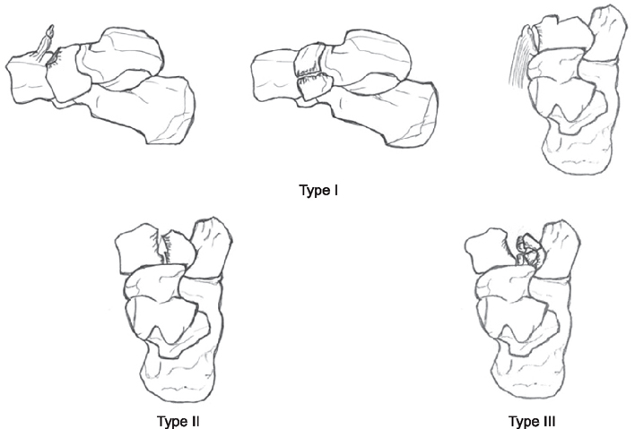
Fig. 2 Sangeorzan's classification of navicular fracture. Type I: Dorsal or tuberosity Avulsion fracture, the primary fracture line traverse in the coronal plane, which is seen on a lateral radiograph. Type II: Navicular body fracture, in which the fracture line is traverse from dorsolateral to plantarmedial across the body, which is seen on an anteroposterior radiograph. Type III: Navicular body fracture with central or lateral comminution.

Fig. 3 (A) Type II navicular fracture and medial cuneiform subluxation were seen on an oblique radiograph. (B) Navicular fracture fragment and medial cuneiform were reduced and fixed with plate and screws.

Fig. 4 (A) Nutcracker fracture of cuboid was seen on an anteroposterior radiograph. (B) Bridging plate fixation was performed from the calcaneus to the fifth metatarsal bone to restore the length of the lateral column.

Fig. 5 Medial cuneiform fracture with Lisfranc injury was seen on simple radiograph and computed tomography.
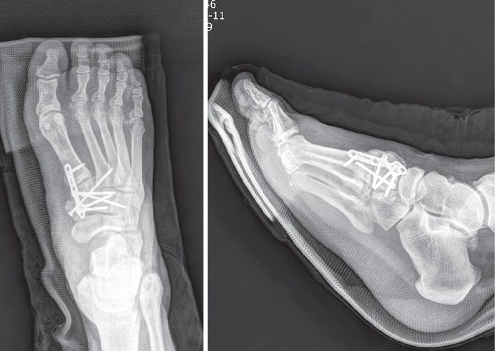
Fig. 6 Fracture of the medial cuneiform and Lisfranc joint were reduced and fixated with plate and screws.
Reference
-
1. Tadros AM, Eid HO, Abu-Zidan FM. Epidemiology of foot injury in a high-income developing country. Injury. 2010; 41:137–140.
Article2. Wei CJ, Tsai WC, Tiu CM, Wu HT, Chiou HJ, Chang CY. Systematic analysis of missed extremity fractures in emergency radiology. Acta Radiol. 2006; 47:710–717.
Article3. Yang GH, Park KC, Yoo GH, et al. Fractures and dislocations of the midfoot and forefoot. In : Yang GH, editor. Principles of fracture management. 1st ed. Seoul: Korean Fracture Society;2013. p. 837–839.4. Lau S, Bozin M, Thillainadesan T. Lisfranc fracture dislocation: a review of a commonly missed injury of the midfoot. Emerg Med J. 2016; DOI: 10.1136/emermed-2015-205317. [Epub].
Article5. Richter M, Wippermann B, Krettek C, Schratt HE, Hufner T, Therman H. Fractures and fracture dislocations of the midfoot: occurrence, causes and long-term results. Foot Ankle Int. 2001; 22:392–398.
Article6. Myerson MS, McGarvey WC, Henderson MR, Hakim J. Morbidity after crush injuries to the foot. J Orthop Trauma. 1994; 8:343–349.
Article7. Thordarson DB. Detecting and treating common fractures of the foot and ankle part 2: the midfoot and forefoot. Phys Sportsmed. 1996; 24:58–64.8. Kadow TR, Siska PA, Evans AR, Sands SS, Tarkin IS. Staged treatment of high energy midfoot fracture dislocations. Foot Ankle Int. 2014; 35:1287–1291.
Article9. DiGiovanni CW. Fractures of the navicular. Foot Ankle Clin. 2004; 9:25–63.
Article10. Sangeorzan BJ, Benirschke SK, Mosca V, Mayo KA, Hansen ST Jr. Displaced intra-articular fractures of the tarsal navicular. J Bone Joint Surg Am. 1989; 71:1504–1510.
Article11. Torg JS, Moyer J, Gaughan JP, Boden BP. Management of tarsal navicular stress fractures: conservative versus surgical treatment: a meta-analysis. Am J Sports Med. 2010; 38:1048–1053.12. Rosenbaum AJ, Uhl RL, DiPreta JA. Acute fractures of the tarsal navicular. Orthopedics. 2014; 37:541–546.
Article13. Simon JP, Van Delm I, Fabry G. Fracture dislocation of the tarsal navicular. Acta Orthop Belg. 1993; 59:222–224.14. Andersen RC, Neiderer K, Martin B, Dancho J. Open reduction-internal fixation of a navicular body fracture with dorsal displacement of the first and second cuneiforms: a case report. J Am Podiatr Med Assoc. 2013; 103:246–249.
Article15. Kim BS, Cho SD, Cho YS, Park TW, So CS. Isolated fracture dislocation of the tarsal navicular: case report. J Korean Soc Fract. 1997; 10:925–928.
Article16. Chu IT, Yoo KH. Isolated dislocation of Tarsal Navicular bone: a case report. J Korean Soc Fract. 1997; 10:86–90.
Article17. Grivas TB, Vasiliadis ED, Koufopoulos G, Polyzois VD, Polyzois DG. Midfoot fractures. Clin Podiatr Med Surg. 2006; 23:323–341.
Article18. Cheng Y, Yang H, Sun Z, Ni L, Zhang H. A rare midfoot injury pattern: navicular-cuneiform and calcaneal-cuboid fracture-dislocation. J Int Med Res. 2012; 40:824–831.
Article19. Hunter JC, Sangeorzan BJ. A nutcracker fracture: cuboid fracture with an associated avulsion fracture of the tarsal navicular. AJR Am J Roentgenol. 1996; 166:888.
Article20. Macciocchi B. Etiology of tarsal cuboid bone fractures. Minerva Chir. 1949; 4:620–622.21. Sangeorzan BJ, Swiontkowski MF. Displaced fractures of the cuboid. J Bone Joint Surg Br. 1990; 72:376–378.
Article22. Borrelli J Jr, De S, VanPelt M. Fracture of the cuboid. J Am Acad Orthop Surg. 2012; 20:472–477.
Article23. Newell SG, Woodle A. Cuboid syndrome. Phys Sportsmed. 1981; 9:71–76.
Article24. Patterson SM. Cuboid syndrome: a review of the literature. J Sports Sci Med. 2006; 5:597–606.25. Patterson RH, Petersen D, Cunningham R. Isolated fracture of the medial cuneiform. J Orthop Trauma. 1993; 7:94–95.
Article26. Miersch D, Wild M, Jungbluth P, Betsch M, Windolf J, Hakimi M. A transcuneiform fracture-dislocation of the midfoot. Foot (Edinb). 2011; 21:45–47.
Article27. Lee BH, Shin DM. Treatment of fracture - dislocation of Tarsometatarsal joint. J Korean Soc Fract. 1995; 8:606–614.
Article28. Ahn SH, Kim HC, Kim KY, Yoon HJ, Kim IY. Avulsion fracture of medial cuneiform by Tibialis anterior tendon (a case report). J Korean Foot Ankle Soc. 2010; 14:194–196.29. Chung HJ, Park JG, Kam MC. Diagnosis and treatment of occult Lisfranc injury. J Korean Foot Ankle Soc. 2013; 17:34–39.30. Khatri Chhetri KM, Acharya P, Rokaya Chhetri DR. Combined fracture dislocation of the navicular bone along with cuboid, cuneiform and longitudinal split fracture of the lateral malleolus: a rare combination of fractures. Chin J Traumatol. 2014; 17:358–360.
- Full Text Links
-
- Actions
-
Cited
- CITED
-
- Close
- Share
-
- Similar articles
-
- A Clinical Study of Peri - tarsal Dislocation
- An Isolated Dislocation of Tarsal Navicular Bone: a Case Report
- Treatment of the Infected Ununited Fractures of the Tibia by Posterior Bone Graft
- Isolated Fracture Dislocation of the Tarsal Navicular: Case report
- Essential Osteolysis of Carpal and Tarsal Bones: A Case Report


