Transvenous Coil Embolization for Dural Arteriovenous Fistulas of the Ophthalmic Sheath: Report of Two Cases and Review of the Literature
- Affiliations
-
- 1Department of Neurosurgery, Samsung Medical Center, Sungkyunkwan University School of Medicine, Seoul, Korea. Jsns.kim@samsung.com
- KMID: 2354890
- DOI: http://doi.org/10.7461/jcen.2016.18.2.135
Abstract
- We present two patients with a dural arteriovenous fistula (dAVF) of the ophthalmic sheath who developed progressive exophthalmos, conjunctival chemosis, and visual loss. These symptoms mimic those of cavernous sinus dAVFs. Dural AVFs of the ophthalmic sheath are extremely rare and their clinical management is controversial. We successfully treated these two patients by transvenous coil embolization. Transvenous embolization appears to be a safe and effective method to treat dAVFs of the ophthalmic sheath.
Keyword
MeSH Terms
Figure
-
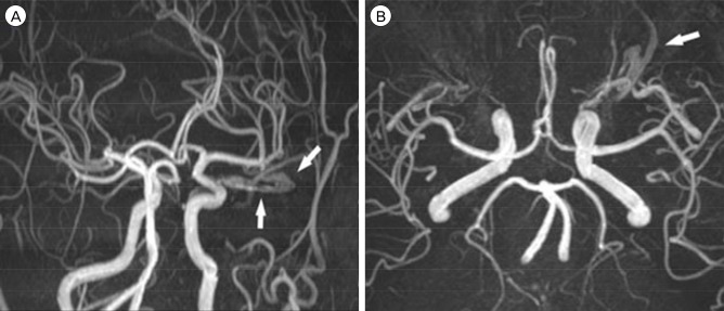
Fig. 1 Frontal (A) and superior (B) 3D TOF MRA maximum-intensity projections show a dilated left superior ophthalmic vein (white arrow). TOF MRA = time-of-flight magnetic resonance angiography.
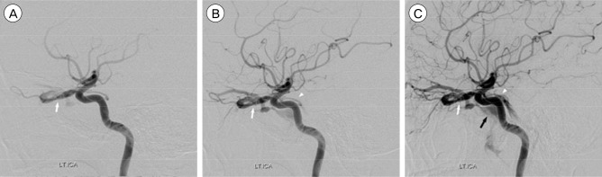
Fig. 2 Lateral projection of left internal carotid angiogram shows dura AVFs of the left optic sheath (white arrow) with fine feeders from the ophthalmic artery and meningohypophyseal trunk (white arrowheads) (A, B). Left optic sheath dural AVF is shown to drain into the cavernous sinus (black arrow) (C). AVF = arteriovenous fistula.
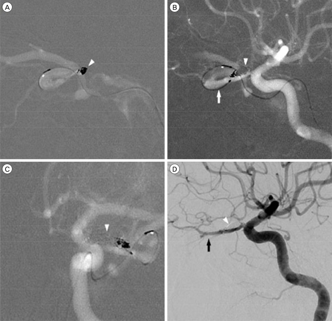
Fig. 3 Angiogram shows the first coil (white arrowhead) inserted into the fistula point of the ophthalmic sheath in lateral view (A) and the fifth coil (white arrowhead) inserted into the shunting point via a transvenous approach. Microcatheter in the superior ophthalmic vein (white arrow) is seen on the lateral view (B) and the AP view (C). Angiogram after embolization shows complete obliteration of left optic sheath dural AVF (white arrowhead) and preserved ophthalmic artery (black arrow) (D). AVF = arteriovenous fistula.
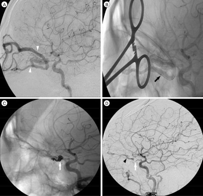
Fig. 4 Angiogram shows dural AVF in the right optic sheath with feeders draining into the superior and inferior ophthalmic veins (white arrowhead) (A). Catheter is directly inserted into the superior ophthalmic vein (black arrow) (B). Coil is inserted into the fistula point of ophthalmic sheath in lateral view (white arrow) (C). Angiogram after embolization shows complete obliteration of right optic sheath dural AVF (white arrow) and preserved ophthalmic artery (black arrowhead) (D). AVF = arteriovenous fistula.
Reference
-
1. Caragine LP Jr, Halbach VV, Dowd CF, Higashida RT. Intraorbital arteriovenous fistulae of the ophthalmic veins treated by transvenous endovascular occlusion: technical case report. Neurosurgery. 2006; 2. 58(1 Suppl):ONS-E170. discussion ONS-E170.2. Cheng KC, Chang CH, Lin WC. Spontaneous resolution of intraorbital arteriovenous fistulas. Ophthal Plast Reconstr Surg. 2009; May-Jun. 25(3):245–247.
Article3. de Keizer R. Carotid-cavernous and orbital arteriovenous fistulas: ocular features, diagnostic and hemodynamic considerations in relation to visual impairment and morbidity. Orbit. 2003; 6. 22(2):121–142. PMID: 12789591.
Article4. Deguchi J, Yamada M, Ogawa R, Kuroiwa T. Transvenous embolization for a purely intraorbital arteriovenous fistula. Case report. J Neurosurg. 2005; 10. 103(4):756–759. PMID: 16266061.5. Hamada J, Morioka M, Kai Y, Sakurama T, Kuratsu J. Spontaneous arteriovenous fistula of the orbit: case report. Surg Neurol. 2006; 1. 65(1):55–57. discussion 57. PMID: 16378859.
Article6. Hieu PD, Besson G, Roncin S, Nonent M. Successful surgical treatment of intraorbital arteriovenous malformations: case report. Neurosurgery. 1997; 3. 40(3):626–631. PMID: 9055307.
Article7. Kim AW, Kosmorsky GS. Arteriovenous communication in the orbit. J Neuroophthalmol. 2000; 3. 20(1):17–19. PMID: 10770500.
Article8. Levy Z, Satti S, Grillo A. Intercavernous carotid artery aneurysm. J Emerg Med. 2014; 2. 46(2):e63–e64. PMID: 24238591.
Article9. Subramanian PS, Gailloud PH, Heck DV, Tamargo RJ, Murphy KJ, Miller NR. Cook detachable coil embolization of a symptomatic, isolated orbital arteriovenous fistula via a superior ophthalmic vein approach. Neuroradiology. 2005; 1. 47(1):62–65. PMID: 15633053.
Article10. Wigton EH, Wells JR, Harrigan MR, Long JA, Vicinanzo MG. Diagnosis and management of an intraorbital vascular fistula. Ophthal Plast Reconstr Surg. 2012; Mar-Apr. 28(2):e39–e41.
Article11. Williamson RW, Ducruet AF, Crowley RW, McDougall CG, Albuquerque FC. Transvenous coil embolization of an intraorbital arteriovenous fistula: case report and review of the literature. Neurosurgery. 2013; 1. 72(1):E130–E134. discussion E134. PMID: 22986598.
- Full Text Links
-
- Actions
-
Cited
- CITED
-
- Close
- Share
-
- Similar articles
-
- Transvenous coil embolization of hypoglossal canal dural arteriovenous fistula using detachable coils: A case report
- Transvenous injection of n-butyl 2-cyanoacrylate to obliterate the pathologic cavernous sinus as a salvage technique for incompletely obliterated complex cavernous sinus dural arteriovenous fistula after transvenous coil embolization
- Middle temporal vein access for transvenous embolization of Cavernous sinus dural arteriovenous fistula: A case report and review of literature
- Transvenous Embolization of Cavernous Sinus Dural Arteriovenous Fistula Using the Direct Superior Ophthalmic Vein Approach: A Case Report
- Facilitated Retrograde Access via the Facial Vein for Transvenous Embolization of the Cavernous Sinus Dural Arteriovenous Fistula with Isolated Ophthalmic Venous Drainage
