Clin Exp Otorhinolaryngol.
2016 Sep;9(3):257-262. 10.21053/ceo.2015.01137.
Histopathologic Evaluations of the Lingual Artery in Healthy Tongue of Adult Cadaver
- Affiliations
-
- 1Department of Otorhinolaryngology-Head and Neck Surgery, Busan St. Mary's Hospital, Busan, Korea.
- 2Department of Pathology, Pusan National University Hospital, Pusan National University School of Medicine, Busan, Korea.
- 3Departement of Otorhinolaryngology-Head and Neck Surgery, Pusan National University School of Medicine, Busan, Korea. wangsg@pusan.ac.kr
- KMID: 2353625
- DOI: http://doi.org/10.21053/ceo.2015.01137
Abstract
OBJECTIVES
To clarify the anatomical distribution of the lingual artery in normal adult subjects through histopathologic evaluations.
METHODS
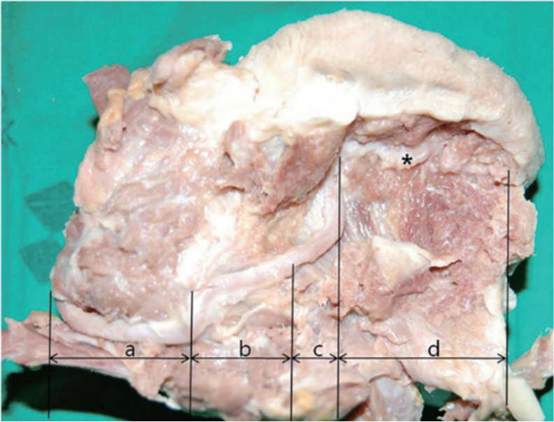
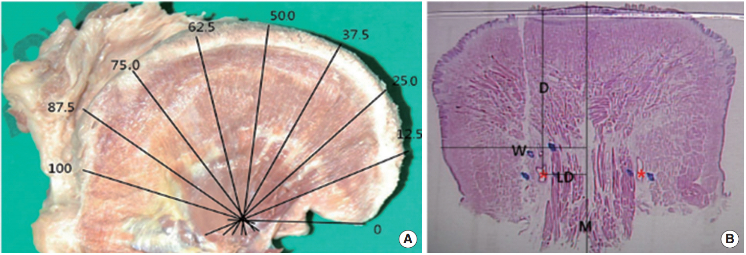
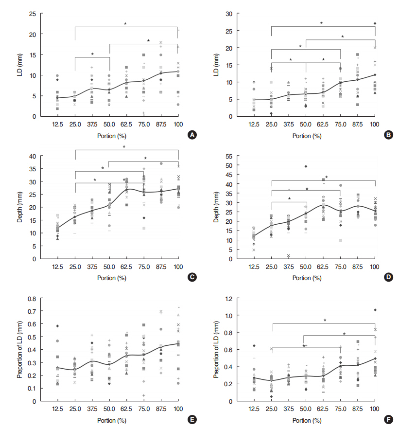
Eighteen healthy cadaveric tongues were used to produce 8 paraffin-embedded tissue sections each. Length from midline raphe, depth from dorsum of tongue and the whole transverse length tongue were measured. The lateral distance, depth, and proportion of lateral distance of deep lingual artery were determined from tip to base of tongue gradually. Lateral distance is length from median raphe to the center of deep lingual artery lumen. Depth is vertical distance from dorsal surface of tongue to the center of deep lingual artery. Proportion of lateral distance is obtained by dividing lateral distance with transverse length from median raphe to lateral border of tongue. The degree of symmetry between right and left sides and the difference between selected spots were evaluated.
RESULTS
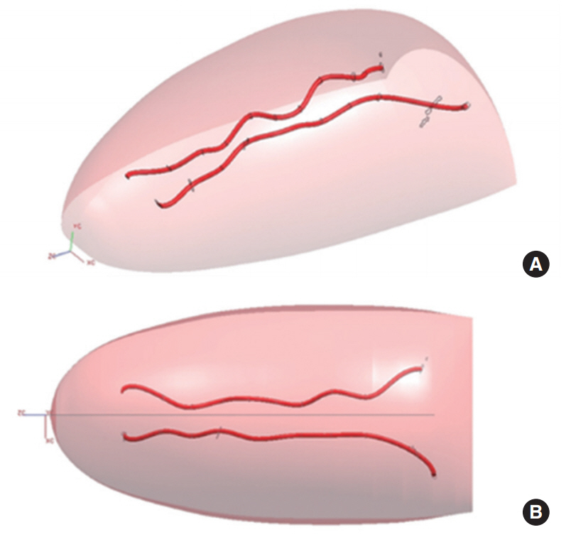
Right and left sides of the lingual artery were symmetric. The lingual artery was lateralized as it run posterior. The lingual artery runs gradually deeper from the surface as it goes near the base of tongue. Both length and depth of the lingual artery gradually increased between 0%-75% of the mobile tongue, but 75%-100% zone of the lingual artery showed no significant difference. There was no anastomosis between right and left side of the lingual arteries. The lingual artery was located within 50% of the transverse length of tongue from median raphe.
CONCLUSION
The present study reveals 3-dimensional information on the anatomical distributions of the lingual artery in normal adult subjects. These findings gives us beneficial information about the handling of the lingual artery during oral and base of tongue-related surgery.
Figure
Reference
-
1. Shangkuan H, Xinghai W, Zengxing W, Shizhen Z, Shiying J, Yishi C. Anatomic bases of tongue flaps. Surg Radiol Anat. 1998; 20(2):83–8.
Article2. Hou T, Shao J, Fang S. The definition of the V zone for the safety space of functional surgery of the tongue. Laryngoscope. 2012; Jan. 122(1):66–70.
Article3. Hou TN, Zhou LN, Hu HJ. Computed tomographic angiography study of the relationship between the lingual artery and lingual markers in patients with obstructive sleep apnoea. Clin Radiol. 2011; Jun. 66(6):526–9.
Article4. Kimura Y, Ariji Y, Gotoh M, Toyoda T, Kato M, Kawamata A, et al. Doppler sonography of the deep lingual artery. Acta Radiol. 2001; May. 42(3):306–11.
Article5. Lopez R, Lauwers F, Paoli JR, Boutault F, Guitard J. Vascular territories of the tongue: anatomical study and clinical applications. Surg Radiol Anat. 2007; Apr. 29(3):239–44.
Article6. Lang P, Thrun B, Hofstetter B, Haberl C, Strehlke P, Hagenmaier C, et al. Giant hemangioma of the tongue: combined use of perioperative blood conservation procedures. Anaesthesist. 2002; Jun. 51(6):470–4.7. Herzog M, Schmidt A, Metz T, Gunthner-Lengsfeld T, Bremert T, Hoppe F, et al. Pseudoaneurysm of the lingual artery after temperature-controlled radiofrequency tongue base reduction: a severe complication. Laryngoscope. 2006; Apr. 116(4):665–7.
Article8. Lins CC, Cavalcanti JS, do Nascimento DL. Extraoral ligature of lingual artery: anatomic and topographic study. Int J Morphol. 2005; 23(3):271–4.
Article9. Houseman ND, Taylor GI, Pan WR. The angiosomes of the head and neck: anatomic study and clinical applications. Plast Reconstr Surg. 2000; Jun. 105(7):2287–313.
Article10. Johnson RE, Sigman JD, Funk GF, Robinson RA, Hoffman HT. Quantification of surgical margin shrinkage in the oral cavity. Head Neck. 1997; Jul. 19(4):281–6.
Article
- Full Text Links
- Actions
-
Cited
- CITED
-
- Close
- Share
- Similar articles
-
- Botulinum Toxin Injection Therapy for Lingual Dystonia: A Case Report
- A Case of Lingual Myoclonus
- Defining Safety Space for Functional Tongue Surgery in Korean Male Obstructive Sleep Apnea Syndrome Patients; Analysis on Spatial Relation of the Tongue and the Lingual Artery
- Lingual Metastasis to the Tip of the Tongue as the First Sign of Metastatic Spread in Lung Cancer: A Case Report and Review of the Literature
- A Case of Non-functioning Lingual Thyroid Excised with COâ‚‚ Laser Via Transoral Approach


