Echocardiographic Evaluation of the Right Heart
- Affiliations
-
- 1Department of Medicine, Virginia Commonwealth University, Richmond, VA, USA. mirasgharali@yahoo.com
- 2Department of Medicine, McGuire VA Medical Center, Richmond, VA, USA.
- KMID: 2353091
- DOI: http://doi.org/10.4250/jcu.2016.24.3.183
Abstract
- The appropriate use of echocardiography may reduce the need for invasive diagnostic cardiac procedures. The right side of the heart has recently gained interest among cardiologists as it became clear that abnormalities of the right heart morphology and function are associated with increased morbidity and mortality. Echocardiography is easy to perform, relatively cheap, readily available and do not pose the risk of ionizing radiation. Conventional 2D and, more recently, 3D echocardiography provides pertinent anatomic and physiologic information about the right side of the heart. Because of the advantages and simplicity of echocardiography it continues to be an excellent tool for evaluating the structure and function of the right side of the heart. This review outlines the uses of echocardiography in evaluating the right heart structure and function.
Keyword
MeSH Terms
Figure
-
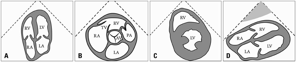
Fig. 1 Echocardiographic views most frequently used to assess the structure and function of the right heart. Apical four chamber view (A). Parasternal short axis view at the level of the aortic valve showing the right atrium (RA), right ventricle (RV), right ventricular outflow tract, pulmonary valve and main pulmonary artery (B). Parasternal short axis view at the level of the papillary muscles (C). Subcostal four chamber view (D). AV: aortic valve, LA: left atrium, LV: left ventricle, PA: pulmonary artery, TV: tricuspid valve.
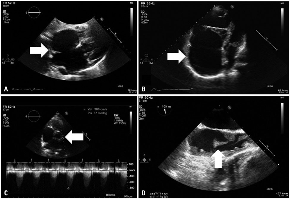
Fig. 2 Echocardiographic images of right heart abnormalities. Atrial septal defect in a 53-year-old woman with transient ischemic attack who declined closure (A, transesophageal echocardiography, 0 degree, arrow pointing at interatrial septum); Ebstein's anomaly in a 41-year-old man with severe tricuspid regurgitation, right atrial enlargement (B, apical four chamber view, arrow showing severely enlarged right atrium), right ventricular dysfunction and right heart failure who subsequently underwent tricuspid valve repair, right atrial reduction and maze procedure; pulmonary valve stenosis in a 38-year-old woman who had congenital pulmonic valve stenosis subsequently corrected by balloon valvuloplasty; Doppler showing high velocities across the valve (C, transesophageal echocardiography, 0 degree, arrow pointing at pulmonary valve plane); and right atrial thrombus (measuring 2 cm in length) in a 30-year-old woman who had systemic lupus erythematosus and end stage renal disease with just removed status of central venous catheter (D, transesophageal echocardiography, 105 degree, arrow pointing at the end of the pedunculated thrombus).
Reference
-
1. Rudski LG, Lai WW, Afilalo J, Hua L, Handschumacher MD, Chandrasekaran K, Solomon SD, Louie EK, Schiller NB. Guidelines for the echocardiographic assessment of the right heart in adults: a report from the American Society of Echocardiography endorsed by the European Association of Echocardiography, a registered branch of the European Society of Cardiology, and the Canadian Society of Echocardiography. J Am Soc Echocardiogr. 2010; 23:685–713. quiz 786-8.2. Napp LC, Luesebrink U, Vogel-Claussen J, Bauersachs J, Roentgen P. Two's company: double-chambered right ventricle [corrected]. Circulation. 2013; 127:e469–e470.3. Jurcut R, Giusca S, La Gerche A, Vasile S, Ginghina C, Voigt JU. The echocardiographic assessment of the right ventricle: what to do in 2010? Eur J Echocardiogr. 2010; 11:81–96. quiz 861-2.4. Valsangiacomo Buechel ER, Mertens LL. Imaging the right heart: the use of integrated multimodality imaging. Eur Heart J. 2012; 33:949–960.5. Horton KD, Meece RW, Hill JC. Assessment of the right ventricle by echocardiography: a primer for cardiac sonographers. J Am Soc Echocardiogr. 2009; 22:776–792. quiz 861-2.6. Jaussi A, Crittin J, Bloch A. [Echocardiography of the right heart--unknown territory. Contribution of echocardiography and Doppler echocardiography to the study of the right heart]. Schweiz Rundsch Med Prax. 1989; 78:494–497.7. Foale R, Nihoyannopoulos P, McKenna W, Kleinebenne A, Nadazdin A, Rowland E, Smith G. Echocardiographic measurement of the normal adult right ventricle. Br Heart J. 1986; 56:33–44.8. Ho SY, Nihoyannopoulos P. Anatomy, echocardiography, and normal right ventricular dimensions. Heart. 2006; 92:Suppl 1. i2–i13.9. Farb A, Burke AP, Virmani R. Anatomy and pathology of the right ventricle (including acquired tricuspid and pulmonic valve disease). Cardiol Clin. 1992; 10:1–21.10. Nesser HJ, Tkalec W, Patel AR, Masani ND, Niel J, Markt B, Pandian NG. Quantitation of right ventricular volumes and ejection fraction by three-dimensional echocardiography in patients: comparison with magnetic resonance imaging and radionuclide ventriculography. Echocardiography. 2006; 23:666–680.11. Bleasdale RA, Frenneaux MP. Prognostic importance of right ventricular dysfunction. Heart. 2002; 88:323–324.12. Kaul S, Tei C, Hopkins JM, Shah PM. Assessment of right ventricular function using two-dimensional echocardiography. Am Heart J. 1984; 107:526–531.13. Pfisterer ME, Battler A, Zaret BL. Range of normal values for left and right ventricular ejection fraction at rest and during exercise assessed by radionuclide angiocardiography. Eur Heart J. 1985; 6:647–655.14. Ghio S, Gavazzi A, Campana C, Inserra C, Klersy C, Sebastiani R, Arbustini E, Recusani F, Tavazzi L. Independent and additive prognostic value of right ventricular systolic function and pulmonary artery pressure in patients with chronic heart failure. J Am Coll Cardiol. 2001; 37:183–188.15. Vonk MC, Sander MH, van den Hoogen FH, van Riel PL, Verheugt FW, van Dijk AP. Right ventricle Tei-index: a tool to increase the accuracy of non-invasive detection of pulmonary arterial hypertension in connective tissue diseases. Eur J Echocardiogr. 2007; 8:317–321.16. Eidem BW, O'Leary PW, Tei C, Seward JB. Usefulness of the myocardial performance index for assessing right ventricular function in congenital heart disease. Am J Cardiol. 2000; 86:654–658.17. Eidem BW, Tei C, O'Leary PW, Cetta F, Seward JB. Nongeometric quantitative assessment of right and left ventricular function: myocardial performance index in normal children and patients with Ebstein anomaly. J Am Soc Echocardiogr. 1998; 11:849–856.18. Hatle L, Sutherland GR. Regional myocardial function--a new approach. Eur Heart J. 2000; 21:1337–1357.19. Isaaz K, Munoz del Romeral L, Lee E, Schiller NB. Quantitation of the motion of the cardiac base in normal subjects by Doppler echocardiography. J Am Soc Echocardiogr. 1993; 6:166–176.20. Yalçin F, Kaftan A, Muderrisoğlu H, Korkmaz ME, Flachskampf F, Garcia M, Thomas JD. Is Doppler tissue velocity during early left ventricular filling preload independent? Heart. 2002; 87:336–339.21. Gondi S, Dokainish H. Right ventricular tissue Doppler and strain imaging: ready for clinical use? Echocardiography. 2007; 24:522–532.22. Papavassiliou DP, Parks WJ, Hopkins KL, Fyfe DA. Three-dimensional echocardiographic measurement of right ventricular volume in children with congenital heart disease validated by magnetic resonance imaging. J Am Soc Echocardiogr. 1998; 11:770–777.23. Jenkins C, Chan J, Bricknell K, Strudwick M, Marwick TH. Reproducibility of right ventricular volumes and ejection fraction using real-time three-dimensional echocardiography: comparison with cardiac MRI. Chest. 2007; 131:1844–1851.24. Gopal AS, Chukwu EO, Iwuchukwu CJ, Katz AS, Toole RS, Schapiro W, Reichek N. Normal values of right ventricular size and function by real-time 3-dimensional echocardiography: comparison with cardiac magnetic resonance imaging. J Am Soc Echocardiogr. 2007; 20:445–455.25. Lu X, Nadvoretskiy V, Bu L, Stolpen A, Ayres N, Pignatelli RH, Kovalchin JP, Grenier M, Klas B, Ge S. Accuracy and reproducibility of real-time three-dimensional echocardiography for assessment of right ventricular volumes and ejection fraction in children. J Am Soc Echocardiogr. 2008; 21:84–89.26. Wang XF, Deng YB, Nanda NC, Deng J, Miller AP, Xie MX. Live three-dimensional echocardiography: imaging principles and clinical application. Echocardiography. 2003; 20:593–604.27. Haddad F, Hunt SA, Rosenthal DN, Murphy DJ. Right ventricular function in cardiovascular disease, part I: anatomy, physiology, aging, and functional assessment of the right ventricle. Circulation. 2008; 117:1436–1448.28. Lindqvist P, Calcutteea A, Henein M. Echocardiography in the assessment of right heart function. Eur J Echocardiogr. 2008; 9:225–234.29. Raymond RJ, Hinderliter AL, Willis PW, Ralph D, Caldwell EJ, Williams W, Ettinger NA, Hill NS, Summer WR, de Boisblanc B, Schwartz T, Koch G, Clayton LM, Jöbsis MM, Crow JW, Long W. Echocardiographic predictors of adverse outcomes in primary pulmonary hypertension. J Am Coll Cardiol. 2002; 39:1214–1219.30. Davlouros PA, Niwa K, Webb G, Gatzoulis MA. The right ventricle in congenital heart disease. Heart. 2006; 92:Suppl 1. i27–i38.31. Tei C, Dujardin KS, Hodge DO, Bailey KR, McGoon MD, Tajik AJ, Seward SB. Doppler echocardiographic index for assessment of global right ventricular function. J Am Soc Echocardiogr. 1996; 9:838–847.32. Park JH, Park MM, Farha S, Sharp J, Lundgrin E, Comhair S, Tang WH, Erzurum SC, Thomas JD. Impaired global right ventricular longitudinal strain predicts long-term adverse outcomes in patients with pulmonary arterial hypertension. J Cardiovasc Ultrasound. 2015; 23:91–99.33. Müller H, Burri H, Lerch R. Evaluation of right atrial size in patients with atrial arrhythmias: comparison of 2D versus real time 3D echocardiography. Echocardiography. 2008; 25:617–623.34. Kilner PJ. Imaging congenital heart disease in adults. Br J Radiol. 2011; 84 Spec No 3:S258–S268.35. Koestenberger M. Transthoracic echocardiography in children and young adults with congenital heart disease. ISRN Pediatr. 2012; 2012:753481.36. Rentzsch A, Abd El Rahman MY, Hui W, Helweg A, Ewert P, Gutberlet M, Lange PE, Berger F, Abdul-Khaliq H. Assessment of myocardial function of the systemic right ventricle in patients with D-transposition of the great arteries after atrial switch operation by tissue Doppler echocardiography. Z Kardiol. 2005; 94:524–531.37. Orie JD, Anderson C, Ettedgui JA, Zuberbuhler JR, Anderson RH. Echocardiographic-morphologic correlations in tricuspid atresia. J Am Coll Cardiol. 1995; 26:750–758.38. Alva C, Ho SY, Lincoln CR, Rigby ML, Wright A, Anderson RH. The nature of the obstructive muscular bundles in double-chambered right ventricle. J Thorac Cardiovasc Surg. 1999; 117:1180–1189.39. Lascano ME, Schaad MS, Moodie DS, Murphy D Jr. Difficulty in diagnosing double-chambered right ventricle in adults. Am J Cardiol. 2001; 88:816–819.40. McElhinney DB, Chatterjee KM, Reddy VM. Double-chambered right ventricle presenting in adulthood. Ann Thorac Surg. 2000; 70:124–127.41. Geibel A. [Echocardiographic evaluation in unoperated congenital heart disease in adults]. Herz. 1999; 24:276–292.42. Fitzgerald KP, Lim MJ. The pulmonary valve. Cardiol Clin. 2011; 29:223–227.43. Hernandez RJ, Bank ER, Shaffer EM, Snider AR, Rosenthal A. Comparative evaluation of the pulmonary arteries in patients with right ventricular outflow tract obstructive lesions. AJR Am J Roentgenol. 1987; 148:1189–1194.44. Yoerger DM, Marcus F, Sherrill D, Calkins H, Towbin JA, Zareba W, Picard MH. Multidisciplinary Study of Right Ventricular Dysplasia Investigators. Echocardiographic findings in patients meeting task force criteria for arrhythmogenic right ventricular dysplasia: new insights from the multidisciplinary study of right ventricular dysplasia. J Am Coll Cardiol. 2005; 45:860–865.45. Kjærgaard J. Assessment of right ventricular systolic function by tissue Doppler echocardiography. Dan Med J. 2012; 59:B4409.46. Apitz C, Webb GD, Redington AN. Tetralogy of Fallot. Lancet. 2009; 374:1462–1471.47. Shinebourne EA, Babu-Narayan SV, Carvalho JS. Tetralogy of Fallot: from fetus to adult. Heart. 2006; 92:1353–1359.48. Bleeker GB, Steendijk P, Holman ER, Yu CM, Breithardt OA, Kaandorp TA, Schalij MJ, van der Wall EE, Nihoyannopoulos P, Bax JJ. Assessing right ventricular function: the role of echocardiography and complementary technologies. Heart. 2006; 92(Suppl 1):i19–i26.49. Dragulescu A, Grosse-Wortmann L, Fackoury C, Mertens L. Echocardiographic assessment of right ventricular volumes: a comparison of different techniques in children after surgical repair of tetralogy of Fallot. Eur Heart J Cardiovasc Imaging. 2012; 13:596–604.50. Warnes CA. Transposition of the great arteries. Circulation. 2006; 114:2699–2709.51. Iriart X, Horovitz A, van Geldorp IE, Barnetche T, Lederlin M, De Guillebon M, Réant P, Lafitte S, Thambo JB. The role of echocardiography in the assessment of right ventricular systolic function in patients with transposition of the great arteries and atrial redirection. Arch Cardiovasc Dis. 2012; 105:432–441.52. George L, Waldman JD, Mathewson JW, Kirkpatrick SE, Pappelbaum SJ, Turner SW. Two-dimensional echocardiographic discrimination of normal from abnormal great artery relationships. Clin Cardiol. 1983; 6:327–332.53. Martin RP, Qureshi SA, Ettedgui JA, Baker EJ, O'Brien BJ, Deverall PB, Yates AK, Maisey MN, Radley-Smith R, Tynan M. An evaluation of right and left ventricular function after anatomical correction and intra-atrial repair operations for complete transposition of the great arteries. Circulation. 1990; 82:808–816.54. Hoffman P, Wójcik AW, Rózański J, Siudalska H, Jakubowska E, Włodarska EK, Kowalski M. The role of echocardiography in diagnosing double chambered right ventricle in adults. Heart. 2004; 90:789–793.55. Choi YJ, Park SW. Characteristics of double-chambered right ventricle in adult patients. Korean J Intern Med. 2010; 25:147–153.56. Reményi B, Wilson N, Steer A, Ferreira B, Kado J, Kumar K, Lawrenson J, Maguire G, Marijon E, Mirabel M, Mocumbi AO, Mota C, Paar J, Saxena A, Scheel J, Stirling J, Viali S, Balekundri VI, Wheaton G, Zühlke L, Carapetis J. World Heart Federation criteria for echocardiographic diagnosis of rheumatic heart disease--an evidence-based guideline. Nat Rev Cardiol. 2012; 9:297–309.57. Pellikka PA, Tajik AJ, Khandheria BK, Seward JB, Callahan JA, Pitot HC, Kvols LK. Carcinoid heart disease. Clinical and echocardiographic spectrum in 74 patients. Circulation. 1993; 87:1188–1196.58. San Román JA, Vilacosta I, López J, Revilla A, Arnold R, Sevilla T, Rollán MJ. Role of transthoracic and transesophageal echocardiography in right-sided endocarditis: one echocardiographic modality does not fit all. J Am Soc Echocardiogr. 2012; 25:807–814.59. Berensztein CS, Piñeiro D, Marcotegui M, Brunoldi R, Blanco MV, Lerman J. Usefulness of echocardiography and doppler echocardiography in endomyocardial fibrosis. J Am Soc Echocardiogr. 2000; 13:385–392.60. Nihoyannopoulos P, Dawson D. Restrictive cardiomyopathies. Eur J Echocardiogr. 2009; 10:iii23–iii33.61. Esmaeilzadeh M, Parsaee M, Maleki M. The role of echocardiography in coronary artery disease and acute myocardial infarction. J Tehran Heart Cent. 2013; 8:1–13.62. Lodato JA, Ward RP, Lang RM. Echocardiographic predictors of pulmonary embolism in patients referred for helical CT. Echocardiography. 2008; 25:584–590.63. Leibowitz D. Role of echocardiography in the diagnosis and treatment of acute pulmonary thromboembolism. J Am Soc Echocardiogr. 2001; 14:921–926.64. Konstantinides S. [Diagnosis and therapy of pulmonary embolism]. Vasa. 2006; 35:135–146.65. Chow BJ, Hassan AH, Chan KL, Tang AS. Prevalence and significance of lead-related thrombi in patients with implantable cardioverter defibrillators. Am J Cardiol. 2003; 91:88–90.66. Eweda II, Samir S, Abbas O, El-Gohary GM, Nammas W. Right heart thrombus-in-transit with pulmonary embolism in a patient with primary hypercoagulable state. Cardiol J. 2010; 17:408–411.67. Meng Q, Lai H, Lima J, Tong W, Qian Y, Lai S. Echocardiographic and pathologic characteristics of primary cardiac tumors: a study of 149 cases. Int J Cardiol. 2002; 84:69–75.68. Gopal AS, Stathopoulos JA, Arora N, Banerjee S, Messineo F. Differential diagnosis of intracavitary tumors obstructing the right ventricular outflow tract. J Am Soc Echocardiogr. 2001; 14:937–940.69. Klarich KW, Enriquez-Sarano M, Gura GM, Edwards WD, Tajik AJ, Seward JB. Papillary fibroelastoma: echocardiographic characteristics for diagnosis and pathologic correlation. J Am Coll Cardiol. 1997; 30:784–790.70. Lacey BW, Lin A. Radiologic evaluation of right ventricular outflow tract myxomas. Tex Heart Inst J. 2013; 40:68–70.71. Sokullu O, Sanioglu S, Deniz H, Ayoglu U, Ozgen A, Bilgen F. Primary cardiac rhabdomyosarcoma of the right atrium: case report. Heart Surg Forum. 2008; 11:E117–E119.72. Amonkar GP, Deshpande JR. Cardiac angiosarcoma. Cardiovasc Pathol. 2006; 15:57–58.73. Labib SB, Schick EC Jr, Isner JM. Obstruction of right ventricular outflow tract caused by intracavitary metastatic disease: analysis of 14 cases. J Am Coll Cardiol. 1992; 19:1664–1668.74. Bodson L, Bouferrache K, Vieillard-Baron A. Cardiac tamponade. Curr Opin Crit Care. 2011; 17:416–424.75. D'Cruz IA, Feghali N, Gross CM. Echocardiographic manifestations of mediastinal masses compressing or encroaching on the heart. Echocardiography. 1994; 11:523–533.
- Full Text Links
-
- Actions
-
Cited
- CITED
-
- Close
- Share
-
- Similar articles
-
- Echocardiographic Findings of Heart Disease in Children
- Echocardiographic Evaluation of Complex Congenital Heart Disease
- Echocardiographic Evaluation of Right-sided Heart in Pediatric Patients with Adenotonsillar Hypertrophy
- Echocardiography in Adult Congenital Heart Diseases
- Echocardiographic parameters and indices in 23 healthy Maltese dogs
