J Korean Ophthalmol Soc.
2016 Sep;57(9):1472-1475. 10.3341/jkos.2016.57.9.1472.
Eyeball Displacement into Ethmoid Sinus with Good Visual Restoration
- Affiliations
-
- 1Department of Ophthalmology, Seoul National University College of Medicine, Seoul, Korea. resourceful@hanmail.net
- 2Department of Ophthalmology, Seoul National University Bundang Hospital, Seongnam, Korea.
- 3Department of Ophthalmology, Seoul Metropolitan Government-Seoul National University Boramae Medical Center, Seoul, Korea.
- KMID: 2351880
- DOI: http://doi.org/10.3341/jkos.2016.57.9.1472
Abstract
- PURPOSE
To report a case of eyeball displacement into the ethmoid sinus followed by early surgical intervention and good visual recovery.
CASE SUMMARY
A 46-year-old female visited our hospital after she injured the right side of her face. Her visual acuity could not be measured and computed tomography revealed displacement of the right eyeball into the ethmoid sinus, as well as right medial orbital wall fracture and rupture of the right medial rectus muscle. She underwent surgical reduction of the herniated eyeball and surgical correction of the medial orbital wall fracture within 20 hours after the accident. Eighteen months after the surgery, visual acuity of the right eye improved from light perception to 20/28, and her color vision and visual field of the right eye improved to normal range.
CONCLUSIONS
Displacement of the eyeball in the orbital wall fracture is very rare, and eyeball displacement into the ethmoid sinus is even rarer. We achieved good visual outcome through early surgical intervention. The early anatomical reduction of the displacement and wall fracture may promote improved final visual outcome in other similar cases.
MeSH Terms
Figure
-
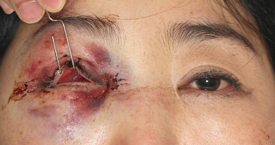
Figure 1. Eyelid photo of the patient. Eyeball could not be found in the palpebral fissure and only conjunctiva was found.

Figure 2. Computed tomography (CT) of the patient. Coronal (A) and axial (B) CT scans showed the medial orbital wall fracture in her right side and the herniated globe (arrow) into the ethmoid sinus.
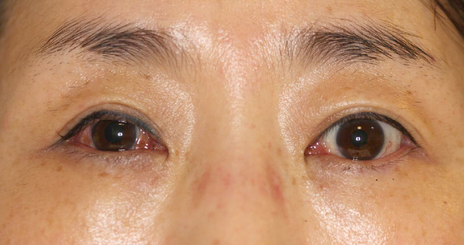
Figure 3. Eyelid photo of the patient 18 months after the eyeball reduction and strabismus surgery. There was only 18 prism diopters of exotropia of the right eye in the primary gaze, despite of the previous rupture of her right medial rectus muscle.
Reference
-
References
1. Pelton RW, Rainey AM, Lee AG. Traumatic subluxation of the globe into the maxillary sinus. AJNR Am J Neuroradiol. 1998; 19:1450–1.2. Haggerty CJ, Roman P. Repositioning of a traumatically displaced globe with maxillary antrostomy: review of the literature and abdominal recommendations. J Oral Maxillofac Surg. 2013; 71:1915–22.3. Okabe H, Kimura K, Sonoda S, Sakamoto T. Displacement of globe into ethmoid sinus by orbital medial wall fracture with good recovery of vision. Jpn J Ophthalmol. 2005; 49:426–8.
Article4. Abrishami M, Aletaha M, Bagheri A, et al. Traumatic subluxation of the globe into the maxillary sinus. Ophthal Plast Reconstr Surg. 2007; 23:156–8.
Article5. Kreiner B, Amer R, Sharfi G, et al. Traumatic displacement of the globe into the paranasal sinuses: case report and guidelines for treatment. J Oral Maxillofac Surg. 2008; 66:826–30.
Article6. Risco JM, Stratas BA, Knott RH. Prolapse of the globe into the ethmoid sinus. Am J Ophthalmol. 1984; 97:659–60.
Article7. Tranfa F, Di Matteo G, Di Salle F, et al. Traumatic displacement of the globe into the ethmoid sinus. Am J Ophthalmol. 2000; 130:253–4.
Article
- Full Text Links
-
- Actions
-
Cited
- CITED
-
- Close
- Share
-
- Similar articles
-
- A Case of Ethmoid Mucocele
- Traumatic Displacement of the Globe into the Ethmoid Sinus: Case Report
- A case of blowout fracture of the orbital wall with eyeball entrapped within the ethmoid sinus
- A Case of Pneumocele in the Ethmoid Sinus
- Correlation of Middle Meatus, Ethmoid Sinus and Maxillary Sinus Microbiology in Patients with Chronic Sinusitis
