Unusual Ultrasonography Findings of Recurred Mammary Fibromatosis Mimicking Subareolar Mastitis: A Case Report
- Affiliations
-
- 1Department of Radiology, College of Medicine, Yeungnam University, Daegu, Korea. ing29@hanmail.net
- 2Department of Pathology, College of Medicine, Yeungnam University, Daegu, Korea.
- KMID: 2349327
- DOI: http://doi.org/10.3348/jksr.2016.75.3.208
Abstract
- Fibromatosis, also known as an extra-abdominal desmoid tumor, rarely occurs in the breast and is often mistaken for carcinoma, clinically and radiologically. Desmoid tumor is a monoclonal myofibroblastic neoplasm which is locally aggressive, but rarely metastasizes. We herein report a case of a 64-year-old woman who experienced two episodes of recurrence of mammary fibromatosis. The mass was initially detected by screening mammography. It appeared as an irregularly shaped mass which was confined within the mammary zone. Recurrences were excised from the right breast 10 and 17 months later. The second recurrence occurred in the subareolar area accompanied by skin thickening and showed an anechoic component on ultrasonography, which mimicked subareolar mastitis with an abscess.
MeSH Terms
Figure
-
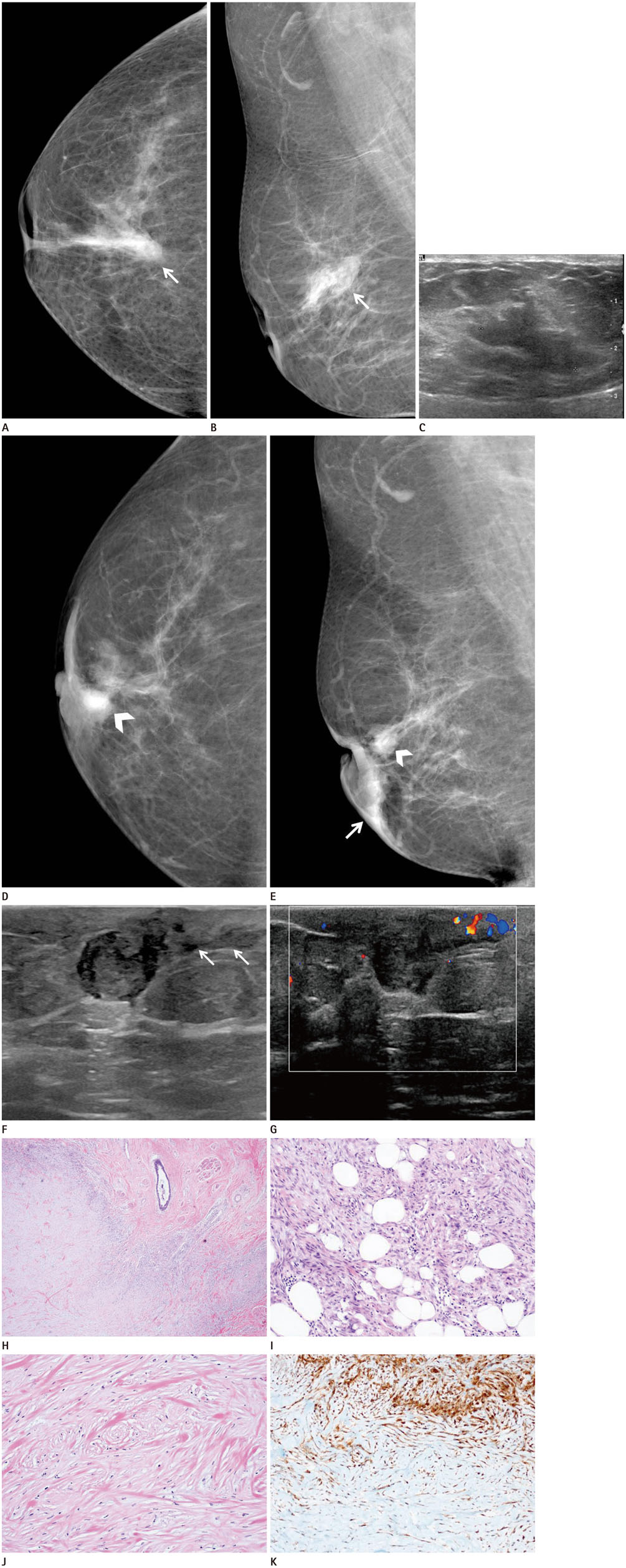
Fig. 1 Radiologic and pathologic findings of recurred mammary fibromatosis in a 64-year-old woman. A, B. Initial presentation of mammary fibromatosis (17 months ago). Craniocaudal (A) and mediolateral oblique views (B) of the Rt. breast show an irregularly shaped, high density mass (arrows) in the subareolar area, confined within the mammary zone. Retraction of the nipple is also seen. C. Ultrasonography demonstrates an irregularly shaped, hypoechoic mass with an indistinct margin in the mammary zone of the right subareolar area. D, E. Radiologic findings of recurred mammary fibromatosis. Craniocaudal (D) and mediolateral oblique views (E) of the Rt. breast show an oval shaped, high density mass (white arrowheads) in the subareolar area with retraction of the nipple and thickening of the overlying skin (white arrow). Calcifications are not seen. F. Ultrasonography of the right breast shows a 1.3 × 0.9 cm sized irregularly shaped mass in the subcutaneous fat layer of the subareolar area. Note, the tubular shaped hypoechoic lesion between the mass and the nipple (white arrows). Increased echogenicity of the surrounding fat tissue and thickening of the overlying skin are also noted. G. Internal vascularity is not observed on color Doppler imaging, but increased vascularity of the surrounding fat tissue and skin is seen. H-K. Photomicrographs of an excised specimen showing fibromatosis. (H) The lesion infiltrates the surrounding breast tissue (H&E, × 40), and (I) the hypercellular peripheral portion shows entrapped fat cells. Proliferation consists of spindle-shaped cells with little or mild cytologic atypia (H&E, × 200). J. The central hypocellular area shows bland cytologic features (H&E, × 200). K. Immunohistochemistry of the excised specimen. The spindle-shaped cells show nuclear and cytoplasmic positivity for beta-catenin immunostain (all × 200). H&E = hematoxylin and eosin
Reference
-
1. Glazebrook KN, Reynolds CA. Mammary fibromatosis. AJR Am J Roentgenol. 2009; 193:856–860.2. Neuman HB, Brogi E, Ebrahim A, Brennan MF, Van Zee KJ. Desmoid tumors (fibromatoses) of the breast: a 25-year experience. Ann Surg Oncol. 2008; 15:274–280.3. Mesurolle B, Ariche-Cohen M, Mignon F, Piron D, Goumot PA. Unusual mammographic and ultrasonographic findings in fibromatosis of the breast. Eur Radiol. 2001; 11:2241–2243.4. Pignatti G, Barbanti-Bròdano G, Ferrari D, Gherlinzoni F, Bertoni F, Bacchini P, et al. Extraabdominal desmoid tumor. A study of 83 cases. Clin Orthop Relat Res. 2000; (375):207–213.5. Wargotz ES, Norris HJ, Austin RM, Enzinger FM. Fibromatosis of the breast. A clinical and pathological study of 28 cases. Am J Surg Pathol. 1987; 11:38–45.6. Rosen PP, Ernsberger D. Mammary fibromatosis. A benign spindle-cell tumor with significant risk for local recurrence. Cancer. 1989; 63:1363–1369.7. Kalbhen CL, Cooper RA, Candel AG. Mammographic and stereotactic core biopsy findings in fibromatosis of the breast: case report. Can Assoc Radiol J. 1998; 49:229–231.8. Jung HK, Kim EK, Ko KH, Kang HY. Breast fibromatosis showing unusual sonographic features. J Ultrasound Med. 2010; 29:1671–1674.9. Lee SM, Lee JY, Lee BH, Kim SY, Joo M, Kim JI. Fibromatosis of the breast mimicking an abscess: case report of unusual sonographic features. Clin Imaging. 2015; 39:685–688.10. Abraham SC, Reynolds C, Lee JH, Montgomery EA, Baisden BL, Krasinskas AM, et al. Fibromatosis of the breast and mutations involving the APC/beta-catenin pathway. Hum Pathol. 2002; 33:39–46.
- Full Text Links
-
- Actions
-
Cited
- CITED
-
- Close
- Share
-
- Similar articles
-
- Mammographic and Sonographic Findings of Periductal Mastitis: A Case Report
- Desmoid-Type Fibromatosis Associated with Silicone Breast Implants
- Autoimmune Mastitis in a Patient with Behcet’s Syndrome: A Case Report with Rapid Changes in Radiologic Features and Characteristic Pathologic Findings
- A Case of Mammary Tuberculosis
- Fine Needle Aspiration Cytology of Periductal Mastitis (Subareolar Abscess) and its Clinical Significance of Cytological Diagnosis
