J Korean Soc Radiol.
2016 Sep;75(3):203-207. 10.3348/jksr.2016.75.3.203.
Descending Colon Endometriosis Misdiagnosed as Diverticulitis: A Case Report
- Affiliations
-
- 1Department of Radiology, Hallym University Sacred Heart Hospital, Hallym University College of Medicine, Anyang, Korea. drkmj@hallym.or.kr
- 2Department of Pathology, Hallym University Sacred Heart Hospital, Hallym University College of Medicine, Anyang, Korea.
- KMID: 2349326
- DOI: http://doi.org/10.3348/jksr.2016.75.3.203
Abstract
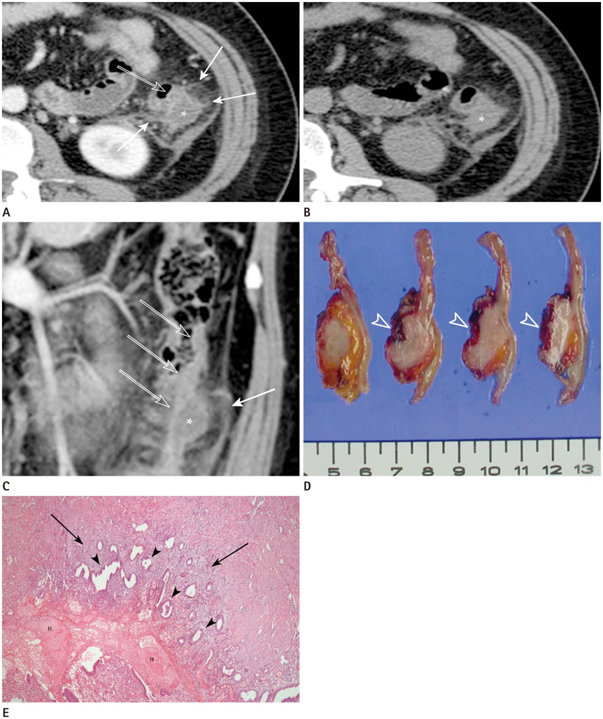
- Endometriosis is defined as the presence of ectopic endometrial tissue outside the uterus. It is a common disease in menstruating females and intestinal involvement is not uncommon. Intestinal endometriosis most commonly involves the sigmoid colon, rectum, ileum, appendix, and cecum. However, the descending colon is a rare site of intestinal endometriosis. Although computed tomography (CT) findings of bowel endometriosis have been presented in several articles, there has been no report describing the CT findings of descending colon endometriosis above the pelvic cavity. Here, we report a rare case of descending colon endometriosis located in the retroperitoneal space, in which the initial impression was acute colonic diverticulitis with a small abscess on preoperative multidetector CT.
MeSH Terms
Figure
Reference
-
1. Garg NK, Bagul NB, Doughan S, Rowe PH. Intestinal endometriosis--a rare cause of colonic perforation. World J Gastroenterol. 2009; 15:612–614.2. Biscaldi E, Ferrero S, Remorgida V, Rollandi GA. Bowel endometriosis: CT-enteroclysis. Abdom Imaging. 2007; 32:441–450.3. Kim JS, Hur H, Min BS, Kim H, Sohn SK, Cho CH, et al. Intestinal endometriosis mimicking carcinoma of rectum and sigmoid colon: a report of five cases. Yonsei Med J. 2009; 50:732–735.4. Yantiss RK, Clement PB, Young RH. Endometriosis of the intestinal tract: a study of 44 cases of a disease that may cause diverse challenges in clinical and pathologic evaluation. Am J Surg Pathol. 2001; 25:445–454.5. Pereira RM, Zanatta A, Preti CD, de Paula FJ, da Motta EL, Serafini PC. Should the gynecologist perform laparoscopic bowel resection to treat endometriosis? Results over 7 years in 168 patients. J Minim Invasive Gynecol. 2009; 16:472–479.6. Remorgida V, Ferrero S, Fulcheri E, Ragni N, Martin DC. Bowel endometriosis: presentation, diagnosis, and treatment. Obstet Gynecol Surv. 2007; 62:461–470.7. Yoon JH, Choi D, Jang KT, Kim CK, Kim H, Lee SJ, et al. Deep rectosigmoid endometriosis: "mushroom cap" sign on T2-weighted MR imaging. Abdom Imaging. 2010; 35:726–731.
- Full Text Links
- Actions
-
Cited
- CITED
-
- Close
- Share
- Similar articles
-
- Colon Cancer Misdiagnosed as Diverticulitis
- A Case of Colon Cancer with Ovarian Metastasis Mimicking Acute Diverticulitis
- Diverticulitis of the Cecum and Ascending Colon
- A stercoral perforation of the descending colon
- Colovesical Fistula Secondary to Sigmoid Diverticulitis Mimicking Bladder Tumor on Ultrasonography: A Case Report


